Download
1 / 1
10 likes | 130 Views
[16:14] And He has pressed into service the things He has created for you in the earth, varying in colors. Surely, in that is a Sign for a people who take heed. B. A non-invasive method of quantifying pancreatic volume in murine models using micro-MRI

E N D
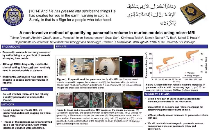
[16:14] And He has pressed into service the things He has created for you in the earth, varying in colors. Surely, in that is a Sign for a people who take heed. B A non-invasive method of quantifying pancreatic volume in murine models using micro-MRI Taimur Ahmad1, Abrahim Orabi1, Jose L. Paredes1, Iman Benbourenane1, Swati Sah1, Kimimasa Tobita2, Sameh Tadros3, Ty Bae3, Sohail Z. Husain1 Departments of Pediatrics1 Developmental Biology2 and Radiology3, Children’s Hospital of Pittsburgh of UPMC & the University of Pittsburgh. BACKGROUND RESULTS • Pancreatic volume is currently assessed by euthanizing a large cohort of animals at varying time points. • Although MRI is frequently used in the clinical setting, it has notbeen routinely used in animal models of disease. • Importantly, nostudies have used MRI imaging to assess pancreas volume in rodents. D C A A Exposed abdomen Figure 1. Preparation of the pancreas for in situ MRI. (A) The peritoneal layer is removed to expose the abdomen and (B) the fixed animal is placed in a conical tube which is inserted in a (C) Bruker 7-tesla micro-MRI. (D) Cross-sectional images are processed in three standard planes. Figure 4. Micro-MRI can reliably measure increases in pancreas volume with increasing age. *, p<0.05 as analyzed using a one way ANOVA. (n=3 per group) AIMS • To test whether micro-MRI can reliably quantify pancreatic volumes in the mouse. D SUMMARY & PLANS B C E SAGITAL 2 CORONAL • MRI is a new part of useful imaging spectrum for mankind, as indicated in the Holy Quran. • Micro-MRI is an accurate and reliable technique for determining pancreatic volume in mice. • MRI can reliably assess increases in pancreatic volume with age. • We will next validate changes in pancreatic volume from murine models of pancreatic injury and obliteration. AXIAL CORONAL SAGITAL METHODS AXIAL • Using a powerful 7-tesla MRI, we performed abdominal imaging on whole-fixed mice. • Traces of the pancreas were transformed into a 3D reconstruction, from which pancreas volumes were generated. Kidney Figure 2. Gross and cross-sectional MRI images of the mouse pancreas. (A) Duodenum, pancreas, and spleen (from left to right) from a mouse. (B-C) Method for generating a 3D reconstruction of the pancreas. (B) The pancreas is traced in each axial section, then cross-checked for accuracy using both (C) sagittal and (D) coronal planes. (E) A 3D reconstruction of the pancreas (in blue) and kidney (in yellow) are generated using these tracings. 3 4 B