Download
1 / 27
470 likes | 1.11k Views
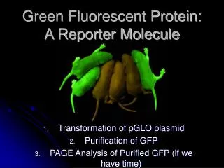
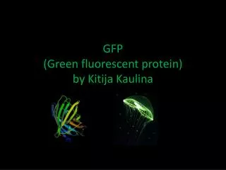
Green Fluorescent Protein. Barzilai Sharon Elissar Naama. Wild Type GFP. GFP traditionally refers to the protein first isolated from the jellyfish Aequorea victoria. GFP is a protein which exhibits bright green fluorescence when exposed to blue light. The protein composed of 238 amino

E N D
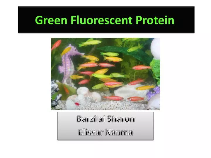
Green Fluorescent Protein Barzilai Sharon ElissarNaama
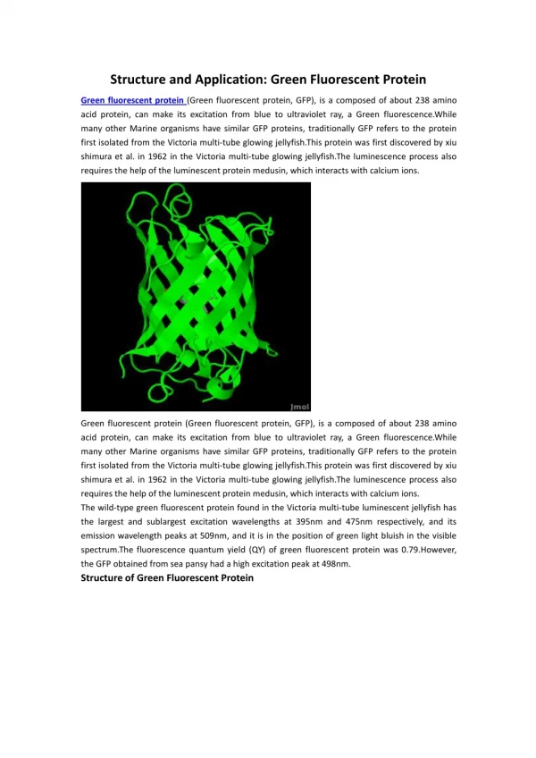
Wild Type GFP • GFP traditionally refers to the protein first isolated from the jellyfish Aequorea victoria. • GFP is a protein which exhibits bright green fluorescence when exposed to blue light. • The protein composed of 238 amino • acids (26.9kDa). • The protein sequence and its resultant • three-dimensional folding structure into • the 11-strand β-barrel, is most likely • crucial to the formation of the • chromophore and its bioluminscene.
Wild Type GFP • The GFP from A. victoria has a major excitation peak at a wavelength of 395 nm and a minor one at 475 nm. Its emission peak is at 509 nm which is in the lower green portion of the visible spectrum. • The GFP from the sea pansy (Renilla reniformis) has a single major excitation peak at 498 nm
The Fluoropore Active Site • GFP has the ability to exhibit intrinsic fluorescence thanks to three amino acids that cyclise (Ser65-Tyr66-Gly67) and then undergo an oxidation step during a complex maturation process. The formed active chromophore contains conjugated double bonds. These double bonds store and release the energy from electrons. The 3 amino acids forming the chormophore in GFP undergoes oxidation and cyclization.
The Fluoropore Active Site • This ability to exhibit intrinsic fluorescence is a major advantage; for • most other fluorescent proteins a cofactor is required (such as ATP) and a • substrates. • Formation is autocatalytic, and the only external requirement is the • presence of oxygen. • http://www.olympusconfocal.com/java/fpfluorophores/gfpfluorophore/index.html
Wild Type GFP blue light is given off by another chemical reaction (bioluminescent or chemiluminescence reaction) involving the protein aequorin inside the jellyfish. This blue light is absorbed by the conjugated double bonds of the chromophore resulting in the release of energy in the form of a visible green light (fluorescent reaction). http://www.lifesci.ucsb.edu/~biolum/chem/index.html
limitations with wild-type GFP • wild-type GFP can be excited by light at two different absorption peaks, which the optimal excitation one is by UV light. This is problematic if one wants to use GFP in live cells, since UV radiation can damage them. • Hence, using techniques such as site-directed mutagenesis, mutants have been created with strong absorption peaks corresponding to blue light.
Limitations with wild-type GFP • Wild-type GFP folds poorly at 37°C, so it limits its usefulness for in vivo experiments in the lab. • mutants of GFP such as Enhanced Green Fluorescent Protein (EGFP) get around this problem. • It can take up to four hours to fold GFP correctly • an engineered version known as Superfolder GFP partly solves this problem.
A smorgasbord of mutants have been derived from GFP to emit different wavelengths of light; these include : • cyan fluorescent protein (CFP) • yellow fluorescent protein (YFP) • blue fluorescent protein (BFP) • The palette of fluorescent proteins available to researchers is supplemented by DsRed, a red fluorescent protein derived from the Discosoma coral, and its derivatives.
Changes in the amino acids in the fluorophore region, as well as in amino acids downstream from the fluorophore, result in changes in the color the fluorophore emits.
Summarizing Green Fluorescent Protein features for applications: • Small: only 26.9kDa in size. • Very stable: has an unusual barrel structure encompassing the hexapeptidechromophore. GFP is resistant to heat, alkaline pH, detergents, photobleaching , chaotropic salts, organic salts, and many proteases. • Fluorescence properties: absorbs light at 395 and 470nm, emits green fluorescence at 509nm. Now altered spectrum available from mutants. • Versatile: could be cloned and expressed in almost all organisms, and every major cellular compartment. • Auto-fluorescence: no fixation needed, no sacrifice of host, no co-factors needed . • Non-invasive and nontoxic in most cases. • Quantifiable: its expression is easily detected, and quantifiable.
Summarizing Green Fluorescent Protein features for applications: • Biological marker . • Fusion of GFP to a protein does not alter the function ,mobility abilities or location of the protein. • This feature has enabled researchers to use GFP in living systems, and it has led to GFP’s widespread use in cell dynamics and development studies.
GFP applications • Reporter Gene: The first applications of GFP were as a reporter gene. • Gene expression can be monitored by using a GFP gene that is under the control of a promoter of interest and measuring the GFP fluorescence which directly indicates the gene expression level in living cells or tissue.
GFP applications • Fusion Tags : A fusion between a cloned gene and GFP. • Insertion of the gene upstream to the GFP-tag results in carboxy-terminal tag on the protein; alternatively, Insertion of the gene downstream from the GFP-tag results in protein with an amino terminal GFP Possible topologies of GFP, cpGFP, and their chimeras with other proteins.
GFP applications FRET (Fluorescence Resonance Energy Transfer): a non-radiative exchange of energy from an excited donor fluorophore to an acceptor fluorophore that is within 10-100 Å from the donor. .
GFP applications Photobleaching: can be used to investigate protein dynamics in living cells.
Specific Examples for the use of GFP Nano Tubes between Macrophages • Showing the creation of Nano-Tubes between human macrophages by the expression of GFP. • Using a bacteria that expresses GFP, the movement of bacteria upon the nano-tube could be seen. B. Onfelt et. al, Structurally Distinct Membrane Nanotubes between Human Macrophages Support Long-Distance Vesicular Traffic or Surfing of Bacteria, The Journal of Immunology, 2006, 177: 8476–8483
Brainbow • A new method that was developed by researchers from the Harvard Brain Center • Brainbow is a genetic “machinery” that randomly mixes green, cyan and yellow fluorescent proteins in individual neurons thereby creating a palette of ninety distinctive hues and colors. • "The technique drives the cell to switch on fluorescent protein genes in neurons more or less at random" Jean Livet
Brainbow • A schematic representation of the method: • In Brainbow transgenes, Cre/lox recombination is used to create a stochastic choice of expression between three or more fluorescent proteins (XFPs)
Brainbow • Integration of tandem Brainbow copies in transgenic mice yielded combinatorial XFP expression, and thus many colours, thereby providing a way to distinguish adjacent neurons and visualize other cellular interactions. • The expression of the XFPs genes was triggered by the injection of Tamoxifen
Brainbow • With a Brainbow construct expressing three XFPs, independent recombination of three transgene copies can, in principle, generate ten distinct colour combinations. J. Livet et al., Transgenic strategies for combinatorial expression of fluorescent proteins in the nervous system, Nature, 2007, 405, p. 56-62
RFP expression in Transgenic Dogs • Fetal fibroblasts were transected with the RFP expressing gene via retroviral vector infection. • The expression of RFP is visible in the transfected fibroblasts as they develop. • Somatic cell nuclear transfer was then employed to replace the nucleus of an oocyte with the nucleus of the RFP-fibroblasts. • The embryos were implanted in the oviducts of surrogate dogs. And…… Six RFP expressing puppies were successfully born!
RFP expression in Transgenic Dogs • In the picture you can the puppy which expresses RFP even under bright field illumination. • The transfection of the RFP expressing gene was also confirmed by southern blot and PCR.
RFP expression in Transgenic Dogs • The organs of the deceased dog express RFP as well. S.G.Hong et al., Generation of Red Fluorescent Protein Transgenic Dogs, genesis, 2008, vol. 47, pg . 314-322
Using GFP in Art Alba the GFP rabbit who initiated a Discussion about the limits between art and science. Austrian artist Gerfried Stocker and molecular biologist Reinhard Nestelbacher grew bacteria in 4,000 Petri-dishes, some of the bacteria expressed GFP and some didn't.
Thank you very much, Have a colorful day!