Download
1 / 42
530 likes | 1.8k Views
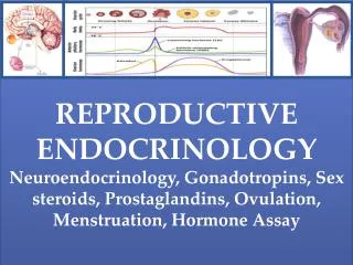
Canine and Feline Endocrinology and Reproductive Anatomy. Dr. N. Matthew Ellinwood, D.V.M., Ph.D. January 27, 2014. Iowa State University College of Agriculture and Life Sciences. Terminology. Endocrinology

E N D
Canine and Feline Endocrinology and Reproductive Anatomy Dr. N. Matthew Ellinwood, D.V.M., Ph.D. January 27, 2014 Iowa State University College of Agriculture and Life Sciences
Terminology • Endocrinology • The study of hormones, the endocrine system, and the role in the physiology of the body. • Endocrine • secreted internally • ductless • applied to organs and structures that release their products into the blood or lymph, and to substances (hormones) that exert specific effects on other organs.
Endocrine System • Functions: • Produce hormones to control other cells, organs or tissues • Major components: • Pancreas • Thyroid • Parathyroid • Adrenal glands • Hypothalamus • Pituitary gland • Gonads • Accessory sex glands www.vrp.com
http://upload.wikimedia.org/wikibooks/en/9/95/Anatomy_and_physiology_of_animals_Main_endocrine_organs_of_the_body.jpghttp://upload.wikimedia.org/wikibooks/en/9/95/Anatomy_and_physiology_of_animals_Main_endocrine_organs_of_the_body.jpg Endocrine Glands of the Dog
Endocrine Systems Major Components • Pancreas • produces insulin and glucagon (endocrine) to regulate blood glucose concentrations • produces digestive enzymes for digestion (exocrine) • Thyroid – produces hormones to regulate mineral concentrations and energy regulation (thyroxine, calcitonin) • Parathyroid – regulates blood Ca 2+ and P+ levels • Adrenal glands • produce steroid hormones • maintain water balance (mineralocorticoids), androgens (sex hormones), and carbohydrate, fat and stress metabolism (glucocorticoids) • produces adrenergic hormones, i.e. stress regulating hormones (norepinephrine, epinephrine)
Endocrine Systems Major Components continued • Hypothalamus • regulates other endrocrine glands (master control) • effects on physiology, emotions, behavior, hunger, etc (GnRH, GHRH, TRH) • Pituitary gland – produces hormones regulating growth, metabolism, reproduction, lactation, and water balance. “The Master Switch” • GH, ACTH, LH, FSH, TSH
Classification of Biochemical Compounds • Almost all biochemical compounds can be classified into four categories • Categories are: • Nucleic acids • Proteins (amino acids and their derivatives) • Lipids • Carbohydrates
http://www.ftmguide.org/images/receptors.gif • Hormones • a chemical substance produced in the body by an organ, cells of an organ, or scattered cells, having a specific regulatory effect on the activity of an organ or organs. • For a hormone to exert its function to the body it must find its specific receptor.
Hormone Categories • Hormones can be classified biochemically, depending on their schemes • Categories are: • Peptides and proteins • Luteinizing hormone (LH) • Amino acid derivatives • Thyroid hormone (T4 and T3) • Steroids • Testosterone, estrogen, and progesterone • Fatty acid derivatives • Prostaglandins
Anatomy of Reproductive Endocrinology • Hypothalamus • Pituitary • Hypothalamo-pituitary axis • Reproductive steroidogenic tissue • Tissue that synthesizes steroids • Testis: Leydig cells • Ovary: ovarian follicle and corpus luteum • Other players • Sertoli cells
Hormones • GnRH • LH • FSH • Testosterone • Progesterone • Estrogen
Feedback Loops • Negative feedback loop • Example: • In the male • Testosterone has a negative feedback effect on GnRH • Positive feedback loop • Example: • In female • Estrogen has a positive feedback effect on GnRH
HYPOTHALAMUS GnRH Hypothalamo-Pituitary Axis ANTERIOR PITUITARY + LH + FSH + Testosterone Sertoli cell Seminiferous tubule Leydig cells of the testis - - Feedback Loops of the Male
HYPOTHALAMUS GnRH Hypothalamo-Pituitary Axis ANTERIOR PITUITARY - LH and FSH Luteal Phase Ovary Follicular Phase Ovary - + - + + LH and FSH + Estrogen + Progesterone Dynamic Feedback Loops of the Female: Depend on Stage of Cycle
Overview of Reproductive Anatomy • Not a class dedicated to Repro anatomy • Will cover basic topics important to basic anatomy and physiology • Specific areas relevant to anatomy and physiology of reproduction in the canine and feline
Fetal Developmental Reproduction • Gonads develop at the caudal pole of the kidney • Female gonad (ovary) remains at this site • Male gonad “migrates” to the scrotum, outside the body wall, via attachment of the gubernaculum • Sperm require lower than body temperature to develop • Failure of normal testicular descent • Cryptorchidism • Unilateral or bilateral • Not monorchid • Mammalian default of the developmental system is female
Anatomy of the Dog and Tom • Testes (plural), testis (singular) • Paired organs, produce male gametes (sperm) and male androgens (testosterone & dihydrotestosterone (DHT)) • Tunica albuginea – dense tough white connective tissue covering testes • Seminiferous tubules, largest structure in testes. Though with a narrow diameter (1/127th of an inch), makes up ~90% of mass of testes • sight of sperm production • Sertoli cells, within seminiferous tubules, aka “nurse cells” or “sustenacular cells” • Leydig cells, in testes interstitiaum (outside seminiferous tubules), hence aka interstitial cells. • Synthesis of testosterone
Cross Section of a Seminiferous Tubule http://openlearn.open.ac.uk/file.php/1638/SK220_1_016i.jpg
Spermatogenesis Rete testis is a central longitudinal collecting duct into which the seminiferous tubules empty. Efferent ductules are located at one pole of the testes and conduct and concentrate immature sperm from the rete testes to the epididymis Epididymis anatomically outside testes (palpable in the dog), site of sperm maturation.
Male Anatomy contin. • Scrotum • sac-like pouch that houses testes, comprised of skin, muscle, and connective tissue • Testes are somewhat moveable within scrotum • Torsions • Cremastor muscles (internal and external - striated) • raise and lower the testes depending on temperature • Tunica dartos muscle (smooth), in scrotal wall • contracts in cold to keep testes warm • Spermatic cord: nerves, vessels, and ductus deferens • Pampiniform plexus is a vascular plexus for counter current heat exchange
Spermatogenesis • Occurs in seminiferous tubules by division of spermatagonium, and supported by Sertoli cells. • Spermatagonium are germ cell stem cells. Divide to repopulate stems cell population (diploid), or to produce sperm (haploid) • Mitotic vs. meiotic division • As spermatids mature, they move from the outside edge of the seminiferous tubule to the lumen.
http://faculty.sunydutchess.edu/Scala/Bio102/PDF/Spermatogenesis.jpghttp://faculty.sunydutchess.edu/Scala/Bio102/PDF/Spermatogenesis.jpg Spermatogenesis
Spermatogenesis • Rete testis is a central longitudinal collecting duct into which the seminiferous tubules empty. • Efferent ductules are located at one pole of the testes and conduct and concentrate immature sperm from the rete testes to the epididymis • Epididymis anatomically outside testes (palpable in the dog), site of sperm maturation.
Sperm Maturation • Epididymis (pl epididymides): critical for normal maturation and function of sperm • Found between efferent ductules and ductus deferens • Caput (head) – absorbs fluid to concentrate sperm • Corpus (body) – maturation of sperm • Cauda (tail) – storage of fertile sperm • Innervated for ejaculation • Epididymitis: inflammation of epididymis, usually the tail. Scarring causes infertility • Brucella canis • Severe inflammation in dog • Males: can cause scrotal necrosis • Females: causes abortions at 40-60 days
Ejaculation • Contractions of smooth muscles of epididymis, ductus etc propel ejaculate • Epididymis • Ductus (ampulla in dog only) • Abdominal urethra • Prostate gland (both dogs and toms) • Penile urethra
Ejaculate • Semen = sperm + seminal fluid • = ejaculate • Seminal fluid consists of ions (salts), sugars, and buffers. • Activate sperm • Provides nutrients • Provides for stable pH • Provide fluid for lubrication and swimming • Produced by accessory sex glands
Male Accessory Sex Glands • Prostate (dog and tom) • Clinically important in disease in the dog • Prostatic abscess • Bulbourethral gland (tom only), cleanses urethra prior to ejaculation • Seminal vesicles (absent in both)
http://www.goddardvetgroup.co.uk/images/anatomy_physiology_6.jpghttp://www.goddardvetgroup.co.uk/images/anatomy_physiology_6.jpg
http://instruction.cvhs.okstate.edu/Histology/images/10Xpenis3.jpghttp://instruction.cvhs.okstate.edu/Histology/images/10Xpenis3.jpg http://upload.wikimedia.org/wikipedia/commons/2/2b/Ospenis.jpg Insemination • Penile anatomy • Cavernosum tissue • os penis
http://www.vivo.colostate.edu/hbooks/pathphys/reprod/semeneval/dogcoll3.jpghttp://www.vivo.colostate.edu/hbooks/pathphys/reprod/semeneval/dogcoll3.jpg • Bulbis glandis (dog only) • Responsible for the tie or coital lock • Penile spines (tom only) • Responsible for inducing ovulation • Dependent on testosterone production http://www.vetmed.lsu.edu/eiltslotus/theriogenology-5361/cat%20penis%20spines.jpg
http://www.nature.com/nrg/journal/v4/n12/images/nrg1225-f1.jpghttp://www.nature.com/nrg/journal/v4/n12/images/nrg1225-f1.jpg Female Reproductive Anatomy • Developmental aspects. • Gonads (ovaries) remain at caudal pole of the kidneys • Anatomy results from degree of fusion of Mullerian Ducts, e.g. marsupials
Canine and feline • Bipartite or bicornuate uterus • Well developed uterine horns, single short uterine body, single cervix, single vagina www.webmm.ahrq.gov/.../images/case18_fig1.jpg
Female Gamete Production • Ovaries • Produce female gametes (ovum, ova plural) • Oocytes produced in follicles which develop in waves • Primordial, primary, secondary, antral, pre-ovulatory • Early stages, gonadotrope independent • Independent of gonadotrophs due to the lack of hormone receptors • Later stages, gonadotrope dependent • Dependent of gonadotrophs due to hormone receptors being present.
Follicular Development http://openlearn.open.ac.uk/file.php/1638/SK220_1_017i.jpg
Ovulation • Multiple follicles develop on each ovary • Canine can have two oocytes per follicle • Ovulation and follicular fate • Preovulatory follicle • Corpora hemorrhagicum • Corpora Luteum • Luteolysis • Corpora Albicans • Canine ovulates a primary oocyte • Unusually immature
Fertilization Of Oocyte • Ovarian bursa • Oviduct (one to each ovary) • Site of oocyte maturation and fertilization • Three anatomical and physiological distinct elements • Infundibulum (small in bitches and queens) • Ampulla • Isthmus • “Connects” ovary to uterus
Uterus • Tubular structure which supports the fetus through the placenta • Vaginal bands or strictures • A muscular component (smooth) and an epithelial component • Myometrium (muscular) • Endometrium (epithelial) • Ectopic pregnancy in the bitch and queen • VERY RARE: could be due to ovarian bursa • One report in queen
http://animalsciences.missouri.edu/reprod/Notes/comp-cat/Image86.gifhttp://animalsciences.missouri.edu/reprod/Notes/comp-cat/Image86.gif Insemination/Fertilization • External anatomy • Vulva • Internal • Vestibule • Urinary papilla • Clitoris • Vagina • Cervix
http://www.vivo.colostate.edu/hbooks/pathphys/reprod/placenta/catplac2.jpghttp://www.vivo.colostate.edu/hbooks/pathphys/reprod/placenta/catplac2.jpg Migration and Implantation • Sperm deposited in vagina, and via swimming and contractions come to anterior aspect of uterine horn. • Fertilization in oviduct • Developing embryos • Migrate within uterus • Migrate between horns • Evenly spaced • Implantation via zonary placenta