Download
1 / 26
260 likes | 372 Views
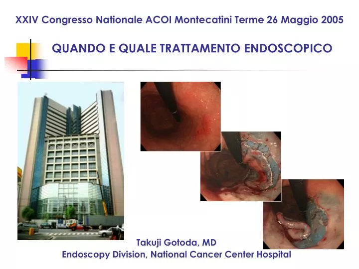
QUANDO E QUALE TRATTAMENTO ENDOSCOPICO. XXIV Congresso Nationale ACOI Montecatini Terme 26 Maggio 2005. Takuji Gotoda, MD Endoscopy Division, National Cancer Center Hospital. New treatment strategy for early gastric cancer. ●. ●. ●. ●. ●. ●. ●. ●. ●. ●. ●. ●. ●. ●. ●. ●. ●.

E N D
QUANDO E QUALE TRATTAMENTO ENDOSCOPICO XXIV Congresso Nationale ACOI Montecatini Terme 26 Maggio 2005 Takuji Gotoda, MD Endoscopy Division, National Cancer Center Hospital
New treatment strategy for early gastric cancer ● ● ● ● ● ● ● ● ● ● ● ● ● ● ● ● ● cancer ● ● ● ● ● ● ● Endoscopic mucosal resection (EMR) Gastrectomy with lymph node dissection cancer
Rational of endoscopic resection Primary gastric cancer Local disease >Endoscopic resection Local disease >Surgical treatment Lymph nodes Systemic disease >Chemotherapy Peritoneum Blood circulation
Indication : EGC with no risk of LN metastasis Conditions Incidence 95% C.I. Differentiated adenocarcinoma 0/1230 (0%) 0-0.3% Intramucosal cancer No lymph-vascular involvement Irrespective of ulcer findings Tumor less than 3cm Differentiated adenocarcinoma 0/929 (0%) 0-0.4% Intramucosal cancer No lymph-vascular involvement Without ulcer findings Irrespective of tumor size 0/145 (0%) 0-2.5% Differentiated adenocarcinoma Minute submucosal penetration (SM1) No lymph-vascular involvement Tumor less than 3cm Gotoda et al, Gastric Cancer, 2000
Clinical management for patients with EGC Finding EGC Pretreatment evaluation using endoscopy, biopsy, EUS, etc. yes no Endoscopic resection non-curative Histological assessment Surgery (gastrectomy+D2) Recently, LADG, SNS, etc. curative Annual surveillance
c Type 0 IIa+IIc T1 SM ? p Type 0 IIa+IIc T1 M, well differentiated, 30mm, UL(+)
No risk of LN metastasis Conditions Incidence 95% C.I. Differentiated adenocarcinoma 0/1230 (0%) 0-0.3% Intramucosal cancer No lymph-vascular involvement Irrespective of ulcer findings Tumor less than 3cm
Standard EMR procedure Polypectomy; Deyhle et al., Endoscopy, 1973 Strip Biopsy; Tada et al., Gastroenterol Endosc, 1984 EMR-C; Inoue et al., Gastrointest Endosc, 1993 EMR-L; Akiyama et al., Gastrointest Endosc, 1997 Soetikno et al, Gastrointest Endosc, 2003
Endoscopic devices for conventional EMR EMR-L using pneumo-activated EVL device Hard and soft hood for EMR-C
Endoscopic resection by conventional EMR Piecemeal resection One piece resection
Local recurrent gastric cancer after previous EMR Author Methods Recurrence rate Strip Biopsy, EAM 3.5% (15/423) Tanabe et al Strip Biopsy, EMR-C 35.3% (97/266) Kawaguchi et al EMR+Laser 6.7% (11/165) Ida et al EMR 10.9% (21/193) Chonan et al ERHSE 2.3% (8/349) Hirao et al Strip Biopsy 18.2% (54/296) Mitsunaga et al Strip Biopsy 8.5% (53/620) NCCH (1988-1998)
Curability and local recurrence 1987-2003 at NCCH Curative Non-curative Not evaluable One piece(1451) 1194 (82%) 209 (14%) 48 (4%) Local rec. 0 16 8 Piecemeal (331) 148 (45%) 81 (24%) 102 (31%) Local rec. 7 (5%) 26 17
LN metastasis after piecemeal resection 2 years later 3 years later
Histological assessment 1: assess the lateral margin 2: assess submucosal penetration 3: assess lymphatic vascular involvement cut every 2mm
Large one piece resection- by Endoscopic Submucosal Dissection (ESD) - well diff. adenoca., Type 0-IIc, 30x25mm, M, ly0, v0, ul-IIs 20x20mm well diff. adenoca., Type 0-IIc, 8x7mm, M, ly0, v0, ul(-) 50x40mm 65x45mm well diff. adenoca., Type 0-IIc, 21x17mm, M, ly0, v0, ul-IIs
Endoscopic equipments for ESD IT knife Hook knife Flex knife Produced by Olympus Medical Systems Corp.
Curability and local recurrence 1987-2003 at NCCH Curative Non-curative Not evaluable 1194(82%) 209(14%) 48(4%) One piece(1451) 0 16 8 Local rec. 148(45%) 81(24%) 102(31%) Piecemeal (331) 7 (5%) 26 17 Local rec.
Chronological trend of treatment strategyfor patients with early gastric cancer at NCCH 100% Cases 350 Guideline EMR 300 Expanded EMR EMR for patients with major complications 250 Surgery 200 50 150 100 50 ‘99 1988 ‘02 ‘03 1990 1996 ‘00 ‘01
Conclusion EMR provides histological staging ● Curability is confirmed only through histological assessment ● ESD is possible to remove a large en bloc resection ● En bloc makes accurate histological assessment possible, and reduces local recurrences ●
Which way would you choose ? EMR ESD