Download

1 / 89
890 likes | 1.07k Views
Blood – Part 1. Chapter 10. Warm-up: 2/25. What is in blood? What does the Greek term “ cyte ” mean? What is one of the purposes of blood?. Blood. Blood Is the “river of life” that surges within us.

E N D
Blood – Part 1 Chapter 10
Warm-up: 2/25 What is in blood? What does the Greek term “cyte” mean? What is one of the purposes of blood?
Blood • Blood • Is the “river of life” that surges within us. • It transports everything that must be carried from one place to another within the body – nutrients, wastes, and body heat – through blood vessels. • Is the only fluid tissue.
Composition of Blood • Blood contains both solid and liquid components. • Formed Elements – Cellular portion of blood. • Contains living blood cells. • Plasma – The fluid portion of the blood. • Nonliving fluid matrix.
Composition of Blood • If blood is spun in a centrifuge, 3 layers are formed: • Bottom Layer- The heavier formed elements are packed down by centrifugal force. • Most of the reddish mass at the bottom is erythrocytes (RBC that function in O2 transport). • Top Layer - The lighter plasma rises to the top. • Buffy Coat - Thin, whitish layer between the formed elements layer and the plasma layer. • Contains leukocytes (WBC that act in various ways to protect the body) and platelets (cell fragments that function in blood clotting).
Percent Composition of Blood • Percent of the total volume of blood: • Erythrocytes: 45% • Percentage known as hematocrit • WBC and Platelets: <1% • Plasma: 55%
Physical Characteristics of Blood Texture: Sticky opaque fluid; Heavier than water and about 5 times thicker (more viscous) than water Taste: Metallic and salty Color: Varies from scarlet (oxygen-rich) to dull red (oxygen-poor)
Physical Characteristics of Blood pH: Slightly alkaline (pH between 7.35 and 7.45) Temperature – Always slightly higher than body temperature (100.4°F) Body Weight and Volume: Accounts for approximately 8% of body weight; Volume in healthy males is about 5-6 L
Plasma • Plasma – The liquid part of the blood. • Is ~90% water. • Over 100 different substances are dissolved in this straw-colored fluid. • Examples include: nutrients, salts, respiratory gases, hormones, plasma proteins, and various wastes • Functions: • Transports various substances around the body. • Helps to distribute body heat evenly throughout the body.
Plasma Proteins • Plasma Proteins – The most abundant solutes in plasma. • Serve a variety of functions: • Albumin – Contributes to the osmotic pressure of blood, which acts to keep water in the bloodstream. • Clotting Proteins help stop blood loss when a blood vessel is injured. • Antibodies help protect the body from pathogens. • Plasma proteins are NOT taken up by the cells to be used as food fuels or metabolic nutrients.
Composition of Plasma • The composition of blood plasma varies continuously as cells remove or add substances to the blood. • The composition of plasma is kept relatively constant by various homeostatic mechanisms. • When blood proteins drop to undesirable levels: Liver is stimulated to make more proteins. • When the blood starts to become too acidic or too basic: The respiratory system and the kidneys are called into action to restore it
Formed Elements • Make up about 45% of whole blood. • Contains erythrocytes or red blood cells (RBCs). • Function: To ferry O2 to all of the cells of the body. • Contain no organelles. • Lacks both a nucleus and a mitochondria • Make ATP by anaerobic mechanisms, they do not use up any of the O2 they are transporting. • Are literally sacs of hemoglobin (Hb). • Hemoglobin - An iron-containing protein that transports the bulk of the oxygen that is carried by the blood.
Shape of Erythrocytes • Are small cells shaped like biconcave disks. • Flattened discs with depressed centers. • They look like doughnuts when viewed through a microscope. • Their small size and peculiar shape provide a large surface area relative to their volume, making them ideally suited for gas exchange.
Number of Erythrocytes (RBCs) • RBCs outnumber WBCs by about 1000 to 1. • Are the major factor contributing to blood viscosity. • When the number of RBCs increase, blood viscosity increases. • As the number of RBCs decrease, the blood thins and flows more rapidly.
Hemoglobin • The more hemoglobin molecules the RBCs contain, the more O2 they will be able to carry. • Each hemoglobin molecule can bind 4 molecules of O2
Hemoglobin (Hb) • The most accurate way of measuring the oxygen-carrying capacity of the blood is to determine how much Hb it contains. • A single RBC contains ~250 million Hb molecules. • Each Hb molecule can bind 4 molecules of O2. Therefore, each of these tiny RBCs can carry about 1 billion O2 molecules! • The Hb content is slightly higher in men than women.
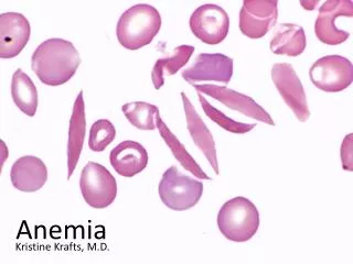
Anemia • Anemia – A decrease in the oxygen-carrying ability of the blood. • May be the result of: • Lower-than-normal number of RBCs • Abnormal or deficient hemoglobin content in the RBCs
Sickle Cell Anemia • Sickle-Cell Anemia – Genetic defect that leads to abnormal Hb, which becomes sharp and sickle-shaped under conditions of increased O2 use by the body. • The deformed (crescent-shaped) RBCs rupture easily and dam up in small blood vessels. • These events interfere with oxygen delivery and cause severe pain.
Sickle Cell Anemia • Occurs chiefly in black people, who live in the malaria belt of Africa and among their descendants. • Individuals with the sickle-cell gene have a better chance of surviving where malaria is prevalent. • The same gene that causes sickle cell anemia, also makes RBCs infected by the malaria-causing parasite stick to the capillary walls and then lose potassium (an essential nutrient for the survival of the parasite). • The malaria causing parasite is prevented from multiplying.
Polycythemia • Polycythemia – An excessive or abnormal increase in the number of erythrocytes. • May result from: • Bone marrow cancer • Normal physiological response to living at high altitudes, where the air is thinner and less oxygen is available. • Major problem that results from high numbers of RBCs is increased blood viscosity. • Increased blood viscosity causes RBCs to flow sluggishly in the body and impairs circulation.
Leukocytes • Leukocytes – White blood cells (WBCs); crucial to the bodies defense against disease. • Far less numerous than RBCs; Account for >1% of total blood volume. • Form a protective, moveable army that helps defend the body against damage by bacteria, viruses, parasites, and tumor cells.
WBCs: Special Characteristics • Are able to slip into and out of the blood vessels – a process called diapedesis. • The circulatory system is simply their means of transportation to areas of the body where their services are needed for inflammatory or immune responses.
WBCs: Special Characteristics • Can locate areas of tissue damage and infection in the body by responding to certain chemicals that diffuse from the damaged cells. • Called positive chemotaxis.
Positive Chemotaxis • Once they have “caught the scent,” the WBCs move through the tissue spaces by ameboid movement . • Ameboid Movement - Flowing cytoplasmic extensions that help them move. • By following the diffusion gradient, they pinpoint the areas of damage and rally round in large numbers to destroy foreign substances or dead cells.
Number of WBCs • Whenever WBCs mobilize for action, the body speeds up their production. • As many as twice the normal number may appear in the blood within a few hours.
Number of WBC’s • Leukocytosis – A total WBC count above 11,000 cells/mm3 • Generally indicates that a bacterial or viral infection is stewing in the body. • Leukopenia – Abnormally low WBC count. • Commonly caused by certain drugs, such as corticosteroids and anticancer agents.
Leukemia • Leukemia – “White blood”; Bone marrow becomes cancerous; Excessive production of abnormal WBCs. • The “newborn” WBCs are immature and incapable of carrying out their normal protective functions. • Consequently, the body becomes easy prey of disease-causing bacteria and viruses.
Classification of WBCs • WBCs are classified into two major groups, depending on whether or not they contain visible granules in their cytoplasm. • Granulocytes – Granule containing WBCs. • Agranulocytes – Lack visible cytoplasmic granules.
Granulocytes • Granulocytes – Granule containing WBCs. • Have lobed nuclei • The granules in their cytoplasm stain specifically with Wright’s stain. • Granulocytes include the: • Neutrophils • Eosinophils • Basophils
Neutrophils • Multilobed nucleus. • Very fine granules that respond to both acid and basic stains. • The cytoplasm as a whole stains pink. • Avid phagocytes at sites of acute infection.
Eosinophils • Blue-red nucleus. • Bilobed- Resembles an old fashioned telephone receiver. • Large brick-red cytoplasmic granules. • Their number increases rapidly during allergies and infections by parasitic worms (ex: tapeworms and flatworms)
Basophils • Rarest of the WBCs. • U-shaped nucleus • Contain large histamine- containing granules that stain dark blue. • Histamine – Inflammatory chemical that makes blood vessels leaky and attracts other WBCs to the inflammatory site.
Agranulocytes • Agranulocytes – lack visible cytoplasmic granules. • Their nuclei is closer to the norm – that is spherical, oval, or kidney shaped. • The agranulocytes include: • Lymphocytes • Monocytes
Lymphocytes • Have a large, dark purple nucleus that occupies most of the cell volume. • Slightly larger than RBCs. • Tend to take up residence in lymphatic tissues, where they play an important role in the immune response.
Monocytes • Largest of the WBCs. • Except for their more abundant cytoplasm and indented nucleus, they resemble large lymphocytes. • When they migrate into the tissues, they change into macrophages with huge appetites. • Macrophages are very important in fighting chronic infections.
Platelets • Platelets – One of the irregular cell frag- ments of blood. • Not cells in the strict sense; They are fragments of bizarre multinucleate cells called megakaryocytes. • Megakaryocytes rupture releasing thousands of anucleate “pieces” that quickly seal themselves off from the surrounding fluids.
Platelets • Appear as darkly staining, irregularly shaped bodies scattered among the other blood cells. • Needed for the clotting process.
Hematopoiesis • Hematopoiesis – Blood cell formation. • Occurs in red bone marrow. • This tissue is found chiefly in the: • Flat bones of the skull and pelvis • Ribs • Sternum • Proximal epiphyses of the humerus and femur • After the cells mature, they are discharged into the blood vessels surrounding the area.
Hemocytoblast • All the formed elements arise from a common type of stem cell, the hemocytoblast. • Hemocytoblasts resides in the bone marrow. • Their development differs and once a cell is committed to a specific blood pathway it cannot change.
The Aging of RBCs • Because they are anucleate, RBCs are unable to synthesize proteins, grow, or divide. • As they age, RBCs become more rigid and begin to fragment, or fall apart in 100-120 days. • Their remains are eliminated by phagocytes in the liver, spleen, and other body tissues. • Lost cells are replaced by the division of hemocytoblasts.
Developing RBCs • The developing RBCs divide many times and then begin synthesizing huge amounts of Hb. • When enough Hb has been accum- ulated, the nucleus and most organelles are ejected and the cell collapses inward. • The entire develop- mental process from hemocytoblast to mature RBC takes 3-5 days.
Erythropoietin • The rate of erythrocyte production is controlled by a hormone called erythropoietin. • Normally a small amount of erythropoietin circulates in the blood at all times, and RBC are formed at a fairly constant rate. • The kidneys play the major role in producing this hormone. • If blood levels of O2 begin to decline, the kidneys step up their release of erythropoietin. • An excessive amount of O2 in the bloodstream, depresses erythropoietin release.
Formation of Leukocytes and Platelets • The formation of leukocytes is stimulated by two hormones: • Colony Stimulating Factors (CSFs) • Interleukins • The production of platelets is accele- rated by the hormone thrombopoietin.