Download
1 / 15
150 likes | 278 Views
Basics of F unctional M agnetic R esonance I maging. How MRI Works. Put a person inside a big magnetic field Transmit radio waves into the person These "energize" the magnetic field of the H ydrogen nucleus in water ( H 2 O )

E N D
How MRI Works • Put a person inside a big magnetic field • Transmit radio waves into the person • These "energize" the magnetic field of the Hydrogen nucleus in water (H2O) • H2O magnetic energy comes back out as very weak radio waves, which are measured by a radio receiver (RF coil) • Frequency of these radio waves is tuned by changing the magnetic field while they are being received (gradient coil) • Frequency changes let images be created
Basic MRI Concepts - 1 • TR = time between reading data out from same location in the brain • Smaller TR is faster imaging • Slices = images are usually made in thin slices, which must be put together to make up a 3 dimensional volume • It usually takes about 50-100 ms to get the data for one slice image • To cover the whole brain = about 30 slices that are 3 mm thick TR is 1.5 to 3.0 seconds • Slower than the heartbeat; Faster than breathing • Voxel = smallest 3 dimensional unit of imaging
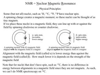
Basic MRI Concepts - 2 • TE = how much time it takes between the radio wave transmit that starts the image, to the center of the image data acquisition • For functional MRI at 3 Tesla, one big problem is image "dropout" (dark regions) in brain regions near air • Nasal sinuses dropout in medial frontal lobe • Ear canals dropout in temporal lobes • Possible solutions (or palliatives): • Thinner slices • Make TE as short as possible
Basic MRI Concepts - 3 • Functional MRI runs MRI scanners very hard • Small problems with the scanner hardware can cause problems with the high speed images that are used for FMRI • Echo Planar Images = EPI • These small problems might not show up in slower images that are used for medical purposes • It is important to check the EPI image quality of your scanner very often by scanning a "phantom" object and looking at the amount of noise • If the noise increases some day, you need help!
D: 4-5 s rise E: 5 s plateau C: 2 s delay B:5 s neural activity F: 4-6 s fall A What is Functional MRI? • 1991: Discovery that MRI-measurable signal increases a few % locally in the brain after increases in neuronal activity (Kwong, et al.) Signal increase caused by change in H2O surroundings: more oxygenated hemoglobin is present =BOLD Cartoon of MRI signal in a single “activated” brain voxel with no noise! G: Return to baseline (or undershoot) A: Pre-activation baseline time
FMRI = It Takes a Team • FMRI is complicated • MRI physics and engineering and operation • Stimulus equipment design and operation • Design of experiment • Analysis of data: AFNI, SPM, FSL, BrainVoyager • Understanding the results of the analysis • FMRI research center needs • MRI physicists or engineers • Statistical experts for data analysis • Computer experts • Plus psychologists and brain scientists!
What Kinds of Questions Can Be Answered with Functional MRI?
Task Based FMRI • To find out information about brain processing of short (1-30 second) stimuli or tasks • Locations in brain that are more or less active in different tasks • and correlations between activation fluctuations • Dependence of neural activation strength (BOLD effect) on task parameters (pain level; face type; …) • Dependence of neural activation on subject parameters (age; disease; …)
Type of Stimuli or Tasks • Short visual or auditory (sound) inputs • Faces / Houses ; Musical tones ; Words • Decision tasks • Same face? Tones up or down? Animal? • You may not care about actual task • You might care about the CONTEXT in which the task appears • Faces: task is MALE or FEMALE but context is angry or fearful face
Groups of Subjects • Can look for differences in activation parameters between group of subjects • Patients and "normals" • Genotypes • Differences in • Activation magnitude • Inter-regional activation correlations • Correlation of activation with covariates
Hard Tasks for FMRI • Anything that requires subject to speak • One word or sound can be OK • Requires censoring out MRI volumes during subject speech — jaw motion is bad for images • Anything that uses subtle sounds (music) • Scanner is very loud • One solution: silent period between scans • Very long duration tasks (learning; drugs) • Hard to tell long activation changes from MRI signal drifting up or down • Not impossible, but requires special analysis
My Advice: Start Small • Do some simple experiment that you KNOW will give results with FMRI • Then increase complexity to get closer to what you really want to do • Do NOT start with your first FMRI experiment being something very complicated and subtle!
FMRI Connectivity • Looking for MRI signal fluctuations that are correlated (vary up and down at same times) in different spatial locations • Can be based on task FMRI or based on "resting" FMRI • Hot new word: Connectome • We have a couple of talks about connectivity analyses in AFNI • Data analysis methods are more variable than for task-based FMRI
Brain "Reading" • Trying to find out what the brain is doing from the FMRI data • Is the subject looking at a face or at an elephant? • Multi-Voxel Pattern Analysis = MVPA • Training data: • To build up different patterns of brain data for different types of brain functions • Support Vector Machines = SVM • Then apply patterns to new brain data to estimate what subject is doing • The limits of MVPA are still being researched