Download
1 / 20
240 likes | 692 Views
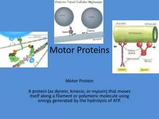
Motor Proteins - Introduction Part 2. Biochemistry 4000 Dr. Ute Kothe. Myosin II. Muscle Myosin ATPase 2 heavy chains (230 kDa): N-terminal globular head + C-terminal long a -helical tail 2 essential light chains (ELC) 2 regulatory light chains (RLC)

E N D
Motor Proteins- Introduction Part 2 Biochemistry 4000 Dr. Ute Kothe
Myosin II • Muscle Myosin • ATPase • 2 heavy chains (230 kDa): N-terminal globular head + C-terminal long a-helical tail • 2 essential light chains (ELC) • 2 regulatory light chains (RLC) • the a-helical tails of 2 heavy chains form a coiled-coil Voet Fig. 35-62
Actin • ATPase • Monomer called G-actin: active site in deep cleft, release of g-phosphates triggers conformational change • polymer called F-actin: double chain of subunits, head-to-tail orientation • (-) end: nucleotide binding cleft • (+) end: opposite end Voet Fig. 35-67 & 35-68
Microfilament Treadmilling • Upon polymerization, F-actin hydrolyzes ATP and releases Pi • Actin-ADP has lower affinity for other actin subunits • Newly polymerized (+) end containing Actin-ATP more stable than (–) end containing Actin-ADP • under steady state, subunits added to (+) end move toward (-) end where they dissociate Voet Fig. 35-80
Structure of Striated Muscle • Thick filaments (purple): myosin II coiled-coil tails packed end to end • Thin filament (gray): F-actin + other proteins (Tropomyosin, Troponin) • Protein-built Z-disk and M-disk which organize and anchor the thick and thin filament Voet Fig. 35-57
Sliding Filament Model • Lenght of thin and thick filaments remains constant • Thick and thin filaments slide past each other • Sliding is driven by many myosin heads (thick filament) walking along F-actin (thin filament) • Overal results in contraction of muscle and generation of force Voet Fig. 35-70
Myosin Cycle • Key features: • ATP reduces Myosin’s affinity for actin • Myosin-ADP strongly binds to actin • Actin binding to Myosin induces phosphate release Voet Fig. 35-71
Myosin Cycle 6. ADP release Actin is ADP release factor • ATP binding • Actin dissociation 2. ATP hydrolysis Cocking of myosin head 5. Power stroke 4. Pi release Strong acting binding 3. Weak actin binding Voet Fig. 35-73
Myosin cycle • ATP binds to Myosin and induces opening of actin binding cleft; Myosin dissociates from actin. • ATPase catalytic site closes and ATP is hydrolyzed inducing a conformational change into high-energy state (cocking); myosin head is moved forward & perpendicular to F-actin • Myosin head binds weakly to actin one monomer further towards Z-disk • Myosin releases phosphate causing the actin binding cleft to close; strengthens myosin-actin interaction • Immediate power stroke: conformational change that sweeps myosin’s C-terminal tail about 10 nm toward the Z-disk relative to the motor domain (head) • ADP is released; actin acts as a nucleotide exchange factor Link to Movie on Bchm4000 webpage!
Myosin II Structure Actin binding cleft Converter domain & Lever Arm Nucleotide Relay Helix Converter domain (green) Lever Arm Voet Fig. 35-62
Myosin versus Kinesin Valle, Science 2000
Conformational changes in Myosin • Presence or absence of g-phosphates influences position of relay helix • Changes in relay helix are transferred to converter domain • Ultimately, result in large displacement of stiff lever arm Voet Fig. 35-74
Is Myosin II a processive motor? • the two myosin heads are not coordinate, cycle independently of each other • net muscle contraction results from uncoordinated actin-attachement and -detachement of many myosins • Myosin II is not processive on its own!
Unconventional Myosin • found in nonmuscle cells: often homodimers, some monomers • mostly move to (+) end of actin, but Myosin VI travels to (-) end • Myosin V: transports cargo via hand-over-hand mechanism, highly processive motor, large step size of net 37 nm per ATP hydrolysis (74 nm movement of one head) Voet Fig. 35-86
Comparison of Myosin & Kinesin Kinesin Myosin Structure Conformational changes Power stroke Step size Processivity
Comparison of Myosin & Kinesin Kinesin Myosin Structure small large same core: ATPase domain, relay helix Conformational changes comparable movement in relay helix different effect on power stroke Power stroke upon ATP binding upon Pi release Step size (per head) 16nm (8nm net) 10nm Processivity highly non-processive
Myosin versus Kinesin Similar structural elements (ATPase domain – blue, relay helix – green, mechanical elements – yellow) Similar conformational changes in Motor domain Power stroke in “different directions” Red/light green: ADP/Nucleotide free Yellow, dark green: ADP-Pi Valle, Science 2000
Model for Power Strokes Myosin Kinesin Power stroke induced by ATP binding Step size: 8 nm Power stroke induced by Pi release Step size: 10 nm Valle, Science 2000
Processivity Kinesin & Myosin V: Highly processive Myosin II: unprocessive Net 37 nm Step size Net 8 nm Step size Valle, Science 2000
Evolution of Motor Proteins Valle, Science 2000