Download
1 / 54
540 likes | 671 Views
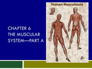
Chapter 7: The Muscle system. Karyn Borrego Tyler Correa Sabrina Cardona Nicholas Jacinto. The three types of muscle. Three types of muscle exist in the human body: smooth, cardiac, and skeletal.

E N D
Chapter 7: The Muscle system Karyn Borrego Tyler Correa Sabrina Cardona Nicholas Jacinto
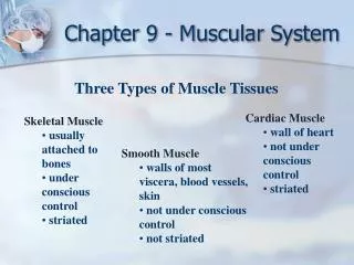
The three types of muscle Three types of muscle exist in the human body: smooth, cardiac, and skeletal. Each fiber is surrounded by a thin layer or areolar connective tissue called endomysium. Smooth: located in the walls of hollow internal organs and blood vessels; moves materials through organs and regulates blood flow in blood vessels; the fibers of this muscle are narrow, tapered root shaped cells; can sustain prolonged contractions and does not fatigue easily; involuntary
Cardiac Muscle • Forms the heart wall. • Fibers are uninucleated , striated, tubular, and branched • Relaxes completely between contractions, which prevents fatigue • Contractions are involuntary
Skeletal muscle • Fibers are tubular, multinucleated, and striated • Contractions always simulated • Supports the body • Makes bones and body parts move, constant body temperature and protection; usually found attached to tendons
In the Laboratory • Muscles can be studied in the lab • When a muscle fiber is isolated and provided with ATP, it contracts completely along its entire length • Result from the all or nothing law: Law that states that muscle fibers contract maximally or not at all • In contrast, a whole muscle shows a degree of contractions • Isolated and stimulated electronically • Mechanical force of contraction is recorded as a visual pattern called myogram
Muscle twitch • Muscle twitch: A single contraction that last for only was fraction of a second • Three parts: • Latent period: period of time between stimulation and initiation of contraction • Contraction period: When the muscle shortens • Relaxation period: When the muscle returns to its former length
Myogram • A. Series of twitches • B. Summation • Increased muscle contraction • C. Tetanic Contraction • Maximal sustained contraction • No longer shows individual twitches; rather the twitches are fused and blended completely into a straight line • Continues until the muscle reaches fatigues: Failure of a muscle fiber to continue to contract due to the exhaustion of ATP
Fatigue • Gradual weakening that occurs after repetitive use • Reasons why muscles become fatigue • ATP is depleted during constants use • Lactic acid by fermentation • Muscles run out of neurotransmitter, acetylcholine • The brain itself signals a person to stop exercising even if the muscles aren’t fatigued
In the body • In the body, muscles are innervated to contract by nerves • Each axon stimulates a number of muscle fibers • Motor Unit: A nerve fiber together with all of the muscle fibers it innervates. • Obeys all-or-none law because the muscles in the motor unit are all stimulated at once; they either contract or do not contract • Tetanic contractions ordinarily occurs in the body because , as the intensity of nervous stimulation increase, more and more motor units are activated • This is known as recruitment • While some muscles are contracting, other are relaxing, which is why muscles rarely experience fatigue • Even when some muscles appear to be resting, they exhibit tone, in which fibers re always contracting. • Muscle tone is important in posture
Exercise and Size of Muscles • Muscles not used or used for weak contraction decrease in size or atrophy. • Atrophy: Occurs when a limb is placed in a cast or when the nerve serving a muscle is damaged. • Can cause muscle fibers to shorten, leaving body parts in contorted positions • If nerve stimulation is not replaced, muscle fibers are replaced by fat & fibrous tissue • Forceful muscular activity causes muscles to increase in size as the number of myofibrils within the muscle fibers • Hypertrophy: occurs only if the muscle contracts to at least 75% of it maximum tension • Athletes take anabolic steroids, either testosterone or related chemicals, to promote muscle growth. This causes undesirable side effects
Slow-twitch muscle fibers • Muscle fibers metabolize both aerobically and anaerobically but some muscle fiber utilizes one method more than the other to provide myofibrils with ATP • Slow-twitch fibers tend to be aerobic and is referred as Type I fibers • Steadier tug and more endurance despite having motor units with a smaller number of fibers • Helpful in sports such as running, biking, jogging • Tire when fuel supply is gone • Have many mitochondria and are dark in color because they contain myoglobin, respiratory pigment found in muscles • Resistant to fatigue because they have a substantial reserve of glycogen and fat
fast twitch muscle fibers • Fast-twitch fibers tend to be anaerobic and is referred as Type II fibers • Designed for strength because their motor units contain many fibers • Provide an explosion of energy and are helpful in sport activities such as sprinting, wrestling and swinging a golf club • Light in color because they have fewer mitochondria, little to no myoglobin, and fewer blood vessels • Develop maximum tension more rapidly than slow twitch fibers and their tension is greater • Vulnerable to an accumulation of lactic acid that causes them to fatigue quickly
Basic Principles • Muscle contracts at a joint, one bone remains stationary and the other moves • The origin of a muscle is on the stationary bone and the insertion of a muscle is on the bone that moves • A body part is moved by a group of muscles working together. A prime mover is the muscle that does most of the work. • The assisting muscles are the synergist • Muscles contract, the shorten. Therefore they can only pull; they cannot push. However, muscles have antagonist. Antagonist pairs work opposite one another to bring about movement in opposite directions
Naming Muscles • Size • Shape • Direction of fibers • Location • Attachment • Number of attachments • Action
Skeletal Muscle Groups The muscles of the body will be grouped according to the region Try to correlate its name with muscles location and action
Muscles of the Head • Facial expression and mastication (chewing) • Neck allows the head to move
Muscles of Facial Expression • Located in scalp and face • Insert into and move the skin • These muscles communicate emotions to others
Muscles of Facial Expressions • Frontalis: Raises the eyebrows • Skin and muscles around the eye • Orbicularis oculi: Closes eye or to blink • Skin around the eye • Orbicularis oris: Closes and protrudes the lips • Skin around the mouth • Buccinator: Compresses cheek inward • Outer surface of maxilla and mandible • Zygomaticus: Raises corner of the mouth • skin and muscle around mouth
Muscles of Mastication • Muscles of mastication are used when we chew and bite • Both Massetersand Temporalis insert on the mandible, are synergist and are prime movers for elevating the mandible • Masseters: Closes jaw • Located in the zygomatic arch • Temporalis: Closes Jaw • Located in the temporal bone • Fan-shaped muscle
Muscles of the Neck • Swallowing • Moving the head
Swallowing • The tongue (a muscle) & the buccinators squeeze the food back along the roof of the mouth to the pharynx • Hyoid: important bone that functions in swallowing , does not articulate with another bone and is attached to the larynx • Suprahyoid Muscles: Lie superior to the Hyoid • Pulls hyoid forward and upward toward the mandible • Infrahyoid Muscles: Lie inferior to the hyoid • Moves the hyoid • Moves hyoid and larynx to their original positions • Epiglottis: Closes the respiratory passages • Small Palatini Muscles: Closes nasal passages • Pharyngeal Constrictor Muscles: Pushes the food into the pharynx; widens the surahyoid muscle
Muscles that move the Head Terms • Flexion: Movement that closes the angle at a joint • Extension: Movement that increases the angle at a joint • Abduction: Movement away from the midline of the body • Adduction: Movement towards the midline of the body • Rotation: Movement of a part around its own axis
Muscles that move the Head • Sternocleidomastoid: Flexes and rotates the head • Located in the sternum and clavicle • Trapezius: Extends the head and adducts the scapula • Located in the Occipital bone C7 vertebra, all of thoracic vertebra
MUSCLES OF THE TRUNK: Thoracic Wall • Primarily involved in breathing • External Intercostal: Elevate rib cage for inspiration during breathing • Located in the superior rib • The Diaphragm: Depress rib cage for forced expiration • Located in the inferior rib • Internal Intercostal: Tenses abdominal wall; lateral rotation of trunk • Located in the lower eight rib
Muscles of the trunk: abdominal wall • Protect and support organs within the abdominal cavity • External: Tenses abdominal wall; lateral rotation of trunk • Located in the lower eight ribs • Internal obliques: Tenses abdominal wall; lateral rotation of trunk • Located in the Iliac crest • Transverse abdominis: Tenses abdominal wall • Located in the lower six • Rectus abdominis: Flexes and rotates the vertebral column • Located in the pubic symphysis
Muscles of the shoulder • The muscles of the shoulder attach the scapula to thorx • Move the scapula • Also attach the humerus to the scapula and move the arm
Muscles that move the scapula • Serratus anterior: Depresses the scapula and pulls it forward; elevates arm above horizontal • Located in the upper nine ribs
Muscles that move the arm • Deltoid: Abducts arm to horizontal • Located in the upper nine ribs • Pectoralis major: Flexes and adducts arms • Located in the spine of scapula and clavicle • Latissimusdorsi: Extends and adducts arm • Located in the lliac crest • Rotator cuff: Angular and rotational movements of arms • Located in the scapula
Muscles that move the forearm • Biceps brachii: Flexes and supinates the forearm • Located in the scapula • Triceps brachii: Extends the forearm • Located in the scapula and proximal humerus • Brachialis: Flexes the forearm • Located in the anterior humerus
MUSCLES THAT MOVE THE HAND AND FINGERS • Flexer carpi and extensor carpi: Move wrist and hand • Located in the humerus • Flexerdigitorum and extensor digitorum: Move fingers • Located in the humerus, radius and ulna
Muscles that move the Thigh • lliposoas: Flexes the thigh • Located in the lumbar vertebrae • Gluteus maximus: Extends the thigh • Located in the Posterior ilium • Gluteus medius: Abducts the thigh • Located in the llium • Adducts group: Adducts the thigh • Located in the Pubis
Muscles that move the leg • Quadriceps femoris group: Extends the legs • Located in the Ilium and femur • Satoruis: Flexes, abducts and rotates leg laterally • Located in the Iiium • Hamstring group: Flexes and rotates leg medially and extends thigh • Located in the Ischial tuberosity