Download
1 / 1
10 likes | 179 Views
Immobilizing the Nematode Caenorhabditis elegans by Addressable Light-Induced Heat Knockdown (ALINK). Han-Sheng Chuang 1,2* , Wen-Tai Chiu 1 and Chang-Shi Chen 3 1 Department of Biomedical Engineering, National Cheng Kung University, Tainan, Taiwan

E N D
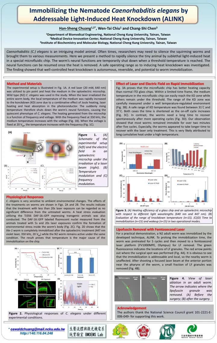
Immobilizing the Nematode Caenorhabditis elegans by Addressable Light-Induced Heat Knockdown (ALINK) Han-Sheng Chuang1,2*, Wen-Tai Chiu1 and Chang-Shi Chen3 1Department of Biomedical Engineering, National Cheng Kung University, Tainan, Taiwan 2Medical Device Innovation Center, National Cheng Kung University, Tainan, Taiwan 3Institute of Biochemistry and Molecular Biology, National Cheng Kung University, Tainan, Taiwan Caenorhabditis (C.) elegans is an intriguing model animal. Often times, researchers may need to silence the squirming worms and brought them to various measurements. Here we present a method to rapidly silence the tiny animal by sublethal light-induced heat in a special microfluidic chip. The worm’s neural functions are temporarily shut down when a threshold temperature is reached. The neural functions can be resumed once the heat is removed. A safe operating range as to inducing heat knockdown was investigated. The finding showed that well-controlled heat knockdown is autonomous, reversible, and potential to worm immobilization. Method and Materials The experimental setup is illustrated in Fig. 1A. A red laser (20 mW, 640 nm) was utilized to pin point and heat the medium in the optoelectric microchip. Wild type (N2) C. elegans was used in the study. When the laser irradiated the entire worm body, the local temperature of the medium was rapidly increased to the konckdown (KD) zone due to a combinative effect of Joule heating, laser heating and heat absorption in the photoconductor. The suddenly rising temperature therefore shuts down the worm’s neural functions, causing the quiescent phenotype of C. elegans. The heating generated from the microchip is a function of frequency and voltage. With the frequency fixed at 350 kHz, the medium temperature increases with the voltage (Fig. 1B). When the voltage is fixed at 20 Vpp, the temperature increases with the frequency (Fig. 1C). Effect of Laser and Electric Field on Rapid Immobilization Fig. 3A proves that the microfluidic chip has better heating capacity than normal ITO glass chips. Within a limited time frame, the medium temperature in the microfluidic chip can easily reach the KD zone while others remain under the threshold. The range of the KD zone was carefully measured under a well temperature-regulated environment (Fig. 3B). A safe range of KD temperature was found between 31°C and 37°C. Both cases the time is shortened as the on-off cycle increases (Fig. 3C). In contrast, the worms need a long time to recover spontaneously after more operating cycles (Fig. 3D). Our observation showed that most worms remained immobile for over two minutes after five cycles. Especially, the worms appeared to take longer time to recover with the laser only treatment. This is very likely attributed to long cumulative heat under a high temperature. Figure 1. (A) Schematic of the experimental setup (left) and the electric field in an optoelectric microchip under the irradiation of a laser beam (right). (B) Temperature modulation and (C) frequency modulation. (B) (C) Physiological Responses C. elegans is very sensitive to ambient environmental changes. The effects of the treatments on worms are shown in Figs. 2A and 2B. The results indicate that the treatment with less than 20s laser exposure can be regarded as no significant difference from the untreated worms. A heat stress evaluation utilizing the TJ356 DAF-16::GFP expressing transgenic animals was also conducted. The DAF-16::GFP labeled fluorescent nuclei measured from the animals treated with 5s and 20s laser exposures confirm the formation of environmental stress inside the worm’s body (Fig. 2C). Fig. 2D shows that the lite-1 worm is completely immobilized after the optoelectric treatment (447 nm violet laser, 350 kHz, 20 Vpp) while the N2 worm remains active under the same condition. The result proves that temperature is the major cause of the immobilization on the chip. Figure 3. (A) Heating efficiency of a glass chip and an optoelectric microchip with respect to different light wavelengths (640 nm and 447 nm). (B) Evaluation of the range of knockdown temperature (n=15). (C)(D) Time to immobilization (n=15) and wakeup (n=15) in two operational modes. Lipofuscin Removal with Femtosecond Laser For a practical demonstration, a N2 adult worm was immobilized by the developed technique, ALINK. To prolong the immobilization time, the worm was pretreated for 5 cycles and then moved to a femtosecond laser platform (FV1000MPE, Olympus) for LF removal. The green fluorescence indicates the locations of LF granules. The red arrow points out where the surgical spot was performed (Fig. 4A). It is obvious to see that the immobilization is addressable and local, so the nearby worm is unaffected. After shooting a focused laser beam at the anterior portion near the pharynx of the worm, a small fraction of LF granules was removed (Fig. 4B). Figure 4. View of laser ablation in an adult worm. The arrow indicates where the lipofuscin granule was removed. (A) Before the surgery; (B) after the surgery. Acknowledgement The authors thank the National Science Council grant 101-2221-E-006-049- for supporting this work. Figure 2. Physiological responses of C. elegans under different experimental conditions. * oswaldchuang@mail.ncku.edu.tw http://140.116.84.246