Download
1 / 24
260 likes | 459 Views
BME1450 Intro to MRI February 2002. The Basics The Details – Physics The Details – Imaging. Example of MRI Images of the Head. Bone and air are invisible. Fat and marrow are bright. CSF and muscle are dark. Blood vessels are bright. Grey matter is darker than white matter. MRI Imagers.

E N D
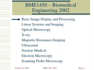
BME1450 Intro to MRIFebruary 2002 The Basics The Details – Physics The Details – Imaging
Example of MRI Images of the Head • Bone and air are invisible. • Fat and marrow are bright. • CSF and muscle are dark. • Blood vessels are bright. • Grey matter is darker than white matter. BME 1450 Introduction to MRI
MRI Imagers GE 1.5 T Signa Imager • GE 0.2T Profile/i imager BME 1450 Introduction to MRI
Basics B1 B0 MR Imaging: Parts of an Imager • Main Magnet • High, constant,Uniform Field, B0. • Gradient Coils • Produce pulsed, linear gradients in this field. • Gx, Gy, & Gz • RF coils • Transmit: B1 Excites NMR signal ( FID). • Receive: Senses FID. B0 B0 BME 1450 Introduction to MRI
Basics RF pulse Gz Gy GX MR Imaging: Pulse Sequence Excitation Excitation Slice Selection A B Phase Encode D Readout E C RF Detected Signal K Space Image Space Coherent detector Complex numbers __________________ __________________ DFT __________________ __________________ __________________ __________________ __________________ ‘Real numbers’ BME 1450 Introduction to MRI
BME595 Intro to MRIOctober 2000 The Basics The Details – Physics The Details – Imaging
The Details - Physics J and Magnetic Resonance (MR) • An object in a magnetic field B0 will become magnetized and develop a net Magnetization, M. • Most of M arises from the orbital electrons but a small part is the Nuclear Magnetization. • The nucleus has a magnetic dipole moment, , and angular momentum,J. • ||/|J| = , the gyromagnetic ratio. • For Hydrogen = 43 MHz/T. Magnetization is “magnetic dipole moment per unit volume”. BME 1450 Introduction to MRI
The Details - Physics Z B0 J or Y |B0|••t X MR: Precession • The 1.5T magnetic, B0 field of the MR Imager makes the Hydrogen Nuclei precess around it. • The precession rate,, is the Larmor frequency. • fL = B0 = 43*1.5 = 64MHz for Hydrogen. BME 1450 Introduction to MRI
The Details - Physics Z B0 J or or M Y |B0|••t X MR : Summary • The magnetization,M, is the density of nuclear magnetic dipole moments. • If you tip M away from B0 it will precess, at frequency B0, producing a measurable RF magnetic field. BME 1450 Introduction to MRI
The Details - Physics Z |B1|••t B0 J or or M Y |B0|••t B1 B1 |B0|••t X B0 MR Excitation • You can tip M by applying a circularly polarized RF magnetic field pulse, B1, to the sample. • If B1 is at the Larmor frequency, B0you get this. • M is now precessing about two magnetic fields. • B1 is effective because it tracks M. BME 1450 Introduction to MRI
The Details - Physics M M(t) 0 | | t T 2 Magnitisation Relaxation • The transverse (M) and longitudinal (M||) components of the magnitization change with time. • Two relaxation times T1 (longitudinal) and T2 (transverse). T1 T2 BME 1450 Introduction to MRI
BME595 Intro to MRIOctober 2000 The Basics The Details – Physics The Details – Imaging
The Details - Imaging Magnitisation Relaxation • MRI Contrast is created since different tissues have different T1 and T2. • Gray Matter: (ms) T1= 810, T2= 101 • White Matter: (ms) T1= 680, T2= 92 BME 1450 Introduction to MRI
The Details - Imaging M + V(t) - MR: The FID • As the magnetization precesses it creates its own RF magnetic field. • This field is much smaller than the Exciting RF field. • It can be detected with a standard radio receiver. • The resulting signal from precession is called the FID. Z B0 J or or M Y |B0|••t Lab Frame X How do you maximize the FID? BME 1450 Introduction to MRI
The Details - Imaging M + V(t) - MR: The MR Signal • The FID can be detected by a ‘read out coil’ and amplified in a standard RF amplifier. • It is then input to a coherent detector with two outputs, I and Q. • The detector is phase locked to the excitation pulse. Thus • My’ “In Phase” output, I • Mx’ “Quadrature output, Q = 0 Z MZ M Y’ My’ Rotating Frame X’ BME 1450 Introduction to MRI
Details - Imaging RF pulse Gz Gy GX Gradient Pulses Excitation Excitation { Slice Selection A B Phase Encode D Readout E C RF Detected Signal K Space Image Space Coherent detector Complex numbers __________________ __________________ DFT __________________ __________________ __________________ __________________ __________________ ‘Real numbers’ BME 1450 Introduction to MRI
Details - Imaging I My’ Q Mx’ MRI: The imaging pulses • The phase gradient pulse will cause more precession. • Precession occurs during the readout gradient pulse as well. • During readout I and Q are digitized into a complex value I+jQ and stored in K space. Z MZ M x•Gx Y’ x•Gx t X’ BME 1450 Introduction to MRI
Details - Imaging MRI: Kspace • If kx(t) and ky(t) are defined as shown, then they represent the row and column that the value, digitized at time t, should be assigned to in Kspace BME 1450 Introduction to MRI
Details - Imaging RF pulse Gz Gy A Gx B D E C MRI: Driving through Kspace • times the integral of the Gx(t) and Gy(t) gives the position in Kspace BME 1450 Introduction to MRI
BME595 Intro to MRIOctober 2000 The Basics The Details – Physics The Details – Imaging Details not discussed
The Details - Physics MR: The Rotating Frame • It is much easier to visualize all this if you observe it from a frame of reference which is rotating at the Larmor frequency, fL=B0. • B1 appears motionless in this rotating frame and B0effectively disappears and… • During the excitation pulse, M precesses only about B1 at frequency B1!! Z |B1|••t MZ M Y’ My’ Rotating Frame B1 X’ BME 1450 Introduction to MRI
The Details - Physics MR: The Rotating Frame • When the excitation pulse is over, M is stationary in the rotating frame. • In the Lab frame, however, it is still precessing. Z MZ M Y’ My’ Rotating Frame X’ BME 1450 Introduction to MRI
Details - Imaging zGz B0 MRI – Meaning of “Z Gradient” X Z Y • A “Z gradient” introduces a gradient in the magnetic field in the Z direction. The gradient is produced with resistive coils. • Traditionally the Z gradient is associated with the RF excitation pulse and slice selection. BME 1450 Introduction to MRI
Details - Imaging B0 MRI – Meaning of “X&Y Gradients” X xGx Z Y • An “X or Y gradient” introduces a gradient in the B0 magnetic field in the X or Y direction. • These gradients are traditionally associated with readout and phase encode, respectively. BME 1450 Introduction to MRI