Download
1 / 10
120 likes | 415 Views
Polycythemia Vera. Aswad H. Al.Obeidy FICMS, FICMS GE&Hep Kirkuk General Hospital. Polycythemia Vera. The World Health Organization (WHO) classification of the chronic myeloproliferative diseases includes seven disorders

E N D
Polycythemia Vera Aswad H. Al.Obeidy FICMS, FICMS GE&Hep Kirkuk General Hospital
Polycythemia Vera • The World Health Organization (WHO) classification of the chronic myeloproliferative diseases includes seven disorders • Chronic myelogenous leukemia, [Ph chromosome t(9;22)(q34;11), BCR/ABL-positive] • Chronic neutrophilic leukemia • Chronic eosinophilic leukemia (and the hypereosinophilic syndrome) • Polycythemia vera • Chronic idiopathic myelofibrosis (with extramedullary hematopoiesis) • Essential thrombocythemia • Chronic myeloproliferative disease, unclassifiable
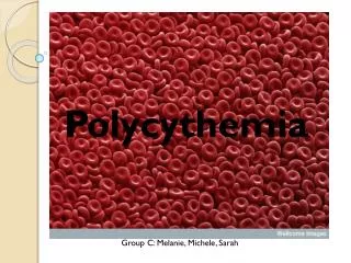
Polycythemia Vera • Polycythemia vera (PV) is a clonal disorder involving a multipotent hematopoietic progenitor cell in which phenotypically normal red cells, granulocytes, and platelets accumulate in the absence of a recognizable physiologic stimulus • The most common of the chronic myeloproliferative disorders, PV occurs in 2 per 100,000 persons, sparing no adult age group and increasing with age to rates as high as 18/100,000 • Familial transmission occurs but is infrequent • A slight overall male predominance has been observed, but women predominate within the reproductive age range
Etiology • The etiology of PV is unknown. Although nonrandom chromosome abnormalities such as 20q, trisomy 8, and especially 9p, have been documented in up to 30% of untreated PV patients, unlike CML no consistent cytogenetic abnormality has been associated with the disorder • However, a mutation in the autoinhibitory, pseudokinase domain of the tyrosine kinase JAK2—which replaces valine with phenylalanine (V617F), causing constitutive activation of the kinase—appears to have a central role in the pathogenesis of PV • JAK2 V617F is the basis for many of the phenotypic and biochemical characteristics of PV, such as elevation of the leukocyte alkaline phosphatase (LAP) score and increased expression of the mRNA of PVR-1, a glycosylphosphatidylinositol (GPI)-linked membrane protein
Etiology • however, JAK2 V617F cannot solely account for the entire PV phenotype. • First, PV patients with the same phenotype and documented clonal disease lack this mutation. • Second, IMF patients have the same mutation but a different clinical phenotype. • Third, familial PV can occur without the mutation, even when other members of the same family express it. • Fourth, not all the cells of the malignant clone express JAK2 V617F. • Fifth, JAK2 V617F has been observed in patients with long-standing idiopathic erythrocytosis.
Clinical Features • Although splenomegaly may be the initial presenting sign in PV, most often the disorder is first recognized by the incidental discovery of a high hemoglobin or hematocrit • With the exception of aquagenic pruritus, no symptoms distinguish PV from other causes of erythrocytosis • Uncontrolled erythrocytosis causes hyperviscosity, leading to neurologic symptoms such as vertigo, tinnitus, headache, visual disturbances, and transient ischemic attacks (TIAs) • Systolic hypertension is also a feature of the red cell mass elevation • In some patients, venous or arterial thrombosis may be the presenting manifestation of PV. Any vessel can be affected, but cerebral, cardiac, or mesenteric vessels are most commonly involved. Intraabdominal venous thrombosis is particularly common in young women and may be catastrophic if a sudden and complete obstruction of the hepatic vein occurs. Indeed, PV should be suspected in any patient who develops hepatic vein thrombosis • Digital ischemia, easy bruising, epistaxis, acid-peptic disease, or gastrointestinal hemorrhage may occur due to vascular stasis or thrombocytosis • Erythema, burning, and pain in the extremities, a symptom complex known as erythromelalgia, is another complication of the thrombocytosis of PV • Given the large turnover of hematopoietic cells, hyperuricemia with secondary gout, uric acid stones, and symptoms due to hypermetabolism
Diagnosis • When PV presents with erythrocytosis in combination with leukocytosis, thrombocytosis, or both, the diagnosis is apparent • However, when patients present with an elevated hemoglobin or hematocrit alone, or with thrombocytosis alone, the diagnostic evaluation is more complex because of the many diagnostic possibilities • Relative erythrocytosis: Hemoconcentration secondary to dehydration, androgens, or tobacco abuse Absolute erythrocytosis • Hypoxia • Carbon monoxide intoxication • High affinity hemoglobin • High altitude • Pulmonary disease • Right-to-left shunts • Sleep-apnea syndrome • Neurologic disease • Renal disease • Renal artery stenosis • Focal sclerosing or membranous glomerulonephritis • Renal transplantation • Tumors • Hypernephroma • Hepatoma • Cerebellar hemangioblastoma • Uterine fibromyoma • Adrenal tumors • Meningioma • Pheochromocytoma • Drugs • Androgens • Recombinant erythropoietin • Familial (with normal hemoglobin function, Chuvash, erythropoietin receptor mutations) • Polycythemia vera
Diagnosis • Once absolute erythrocytosis has been established, its cause must be determined • An elevated plasma erythropoietin level suggests either a hypoxic cause for erythrocytosis or autonomous erythropoietin production, in which case assessment of pulmonary function and an abdominal CT scan to evaluate renal and hepatic anatomy are appropriate • A normal erythropoietin level does not exclude a hypoxic cause for erythrocytosis • In PV, in contrast to hypoxic erythrocytosis, the arterial oxygen saturation is normal. However, a normal oxygen saturation does not exclude a high-affinity hemoglobin as a cause for erythrocytosis; documentation of previous hemoglobin levels and a family study are important • Other laboratory studies that may aid in diagnosis include the red cell count, mean corpuscular volume, and red cell distribution width (RDW) • Only three situations cause microcytic erythrocytosis: -thalassemia trait, hypoxic erythrocytosis, and PV • With -thalassemia trait the RDW is normal, whereas with hypoxic erythrocytosis and PV, the RDW is usually elevated due to iron deficiency • In many patients, the LAP level is also increased, as is the uric acid level. Elevated serum vitamin B12 or B12-binding capacity may be present • In patients with associated acid-peptic disease, occult gastrointestinal bleeding may lead to presentation with hypochromic, microcytic anemia • Unless there is a need to establish the presence of myelofibrosis or exclude some other disorder, bone marrow aspiration need not be done
Complications • The major clinical complications of PV relate directly to the increase in blood viscosity associated with red cell mass elevation and indirectly to the increased turnover of red cells, leukocytes, and platelets with the attendant increase in uric acid and cytokine production • A sudden massive increase in spleen size can be associated with splenic infarction or progressive cachexia • Myelofibrosis appears to be part of the natural history of the disease but is a reactive, reversible process that does not itself impede hematopoiesis and by itself has no prognostic significance • The organomegaly can cause significant mechanical discomfort, portal hypertension, and cachexia • Although the incidence of acute nonlymphocytic leukemia is increased in PV, the incidence of acute leukemia in patients not exposed to chemotherapy or radiation is low • Erythromelalgia is a curious syndrome of unknown etiology associated with thrombocytosis, and is usually responsive to salicylates • Some of the central nervous system symptoms observed in patients with PV, such as ocular migraine, may represent a variant of erythromelalgia • If left uncontrolled, erythrocytosis can lead to thrombosis involving vitalorgans such as the liver, heart, brain, or lungs
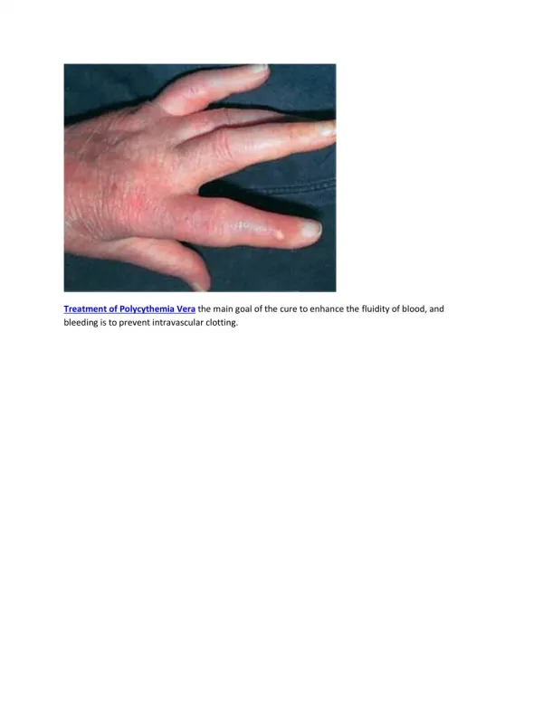
Treatment • PV is generally an indolent disorder whose clinical course is measured in decades • Periodic phlebotomies thereafter serve to maintain the red cell mass within the normal range and to induce a state of iron deficiency, which prevents an accelerated reexpansion of the red cell mass. In most PV patients, once an iron-deficient state is achieved, phlebotomy is usually only required at 3-month intervals • The use of salicylates as a tonic against thrombosis in PV patients is potentially harmful if the red cell mass is not controlled by phlebotomy • Anticoagulants are only indicated when a thrombosis has occurred • Asymptomatic hyperuricemia (<10 mg%) requires no therapy, but allopurinol should be administered to avoid further elevation of the uric acid when chemotherapy is employed to reduce splenomegaly or leukocytosis or to treat pruritus • Generalized pruritus intractable to antihistamines or antidepressants such as doxepin can be a major problem in PV; hydroxyurea, interferon (IFN- ), and psoralens with ultraviolet light in the A range (PUVA) therapy are other methods of palliation • IFN- reduces JAK2 V617F expression in PV patients, and its role in this disorder may be expanding • Anagrelide, a phosphodiesterase inhibitor, can reduce the platelet count and, if tolerated, is preferable to hydroxyurea because it lacks marrow toxicity • Alkylating agents and radioactive sodium phosphate (32P) are leukemogenic in PV, and their use should be avoided • In some patients, massive splenomegaly unresponsive to reduction by hydroxyurea or IFN- therapy and associated with intractable weight loss will require splenectomy • Allogeneic bone marrow transplantation may be curative in young patients • Most patients with PV can live long lives without functional impairment when their red cell mass is effectively managed with phlebotomy. Chemotherapy is never indicated to control the red cell mass unless venous access is inadequate