Download
1 / 1
10 likes | 75 Views
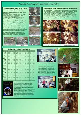
Amphibolite petrography and mineral chemistry. Amphibolites found in the WI/WA areas show three distinct fabrics: Amphibolite bands within mica gneisses of the Tallulah Falls Formation

E N D
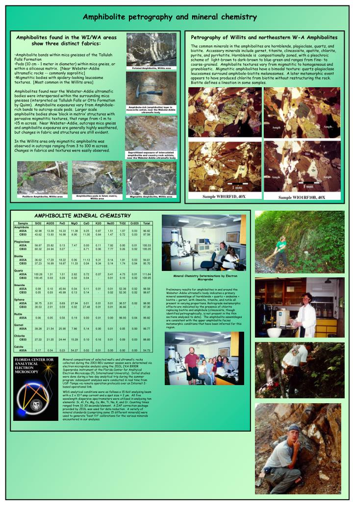
Amphibolite petrography and mineral chemistry • Amphibolites found in the WI/WA areas show three distinct fabrics: • Amphibolite bands within mica gneisses of the Tallulah Falls Formation • Pods (10 cm - 1 meter in diameter) within mica gneiss, or within a siliceous matrix. [Near Webster-Addie ultramafic rocks -- commonly saprolitic] • Migmatitic bodies with spidery-looking leucosome textures. [Most common in the Willits area] • Amphibolites found near the Webster-Addie ultramafic bodies were interspersed within the surrounding mica gneisses (interpreted as Tallulah Falls or Otto Formation by Quinn). Amphibolite exposures vary from Amphibole-rich bands to outcrop-scale pods. Larger scale amphibolite bodies show ‘block in matrix’ structures with pervasive migmatitic textures, that range from <1 m to >15 m across. Near Webster-Addie, outcrops mica gneiss and amphibolite exposures are generally highly weathered, but changes in fabric and structures are still evident. • In the Willits area only migmatitic amphibolite was observed in outcrops ranging from 3 to 100 m across. Changes in fabrics and textures were easily observed. Petrography of Willits and northeastern W-A Amphibolites The common minerals in the amphibolites are hornblende, plagioclase, quartz, and biotite. Accessory minerals include garnet, titanite, clinozoisite, apatite, chlorite, pyrite, and pyrrhotite. Hornblende is compositionally zoned, with a pleochroic scheme of light-brown to dark-brown to blue-green and ranges from fine- to coarse-grained. Amphibolite textures vary from migmatitic to homogeneous and granoblastic. Migmatitic amphibolites have a bimodal texture: quartz-plagioclase leucosomes surround amphibole-biotite melanosomes. A later metamorphic event appears to have produced chlorite from biotite without restructuring the rock. Biotite defines a lineation in some samples. AMPHIBOLITE MINERAL CHEMISTRY Mineral Chemistry Determinations by Electron Microprobe Preliminary results for amphibolites in and around the Webster-Addie ultramafic body indicates a primary mineral assemblage of hornblende + quartz + andesine + biotite ± garnet, with ilmenite, titanite, and rutile all present in varying proportions. Retrograde metamorphic effects are indicated by the presence of chlorite replacing biotite and amphibole (clinozoisite, though identified petrographically, is not present in the thin sections analyzed to date). The amphibolite assemblages are consistent with the upper amphibolite facies metamorphic conditions that have been inferred for this region. Mineral compositions of selected mafic and ultramafic rocks collected during the 2001 REU summer season were determined via electron microprobe analysis using the JEOL JXA 8900R Superprobe instrument at the Florida Center for Analtyical Electron Microscopy (FL International University). Initial studies were done during a two-day analytical trip during the summer program; subsequent analyses were conducted in real time from USF-Tampa via remote operation protocols over an Internet 2-based operational link. WDS analytical conditions were as follows a 15 KeV analyzing beam with a 2 x 10⁻⁸ amp current and a spot size ≈ 2 µm. All five wavelength dispersive spectrometers were utilized in analyzing ten elements: Si, Al, Fe, Mg, Ca, Mn, Ti, Na, K, and Cr. Counting times ranged from 10-30 seconds/element. A ZAF correction package provided by JEOL was used for data reduction. A variety of mineral standards (comprising some 15 different minerals) were used to generate "best fit" calibrations for the various minerals encountered in our analyses.