Download
1 / 1
10 likes | 141 Views
B16 F10 melanoma: an investigation of genetic stability Erica Martin, advised by Dr. Spilatro. Introduction. Conclusions.

E N D
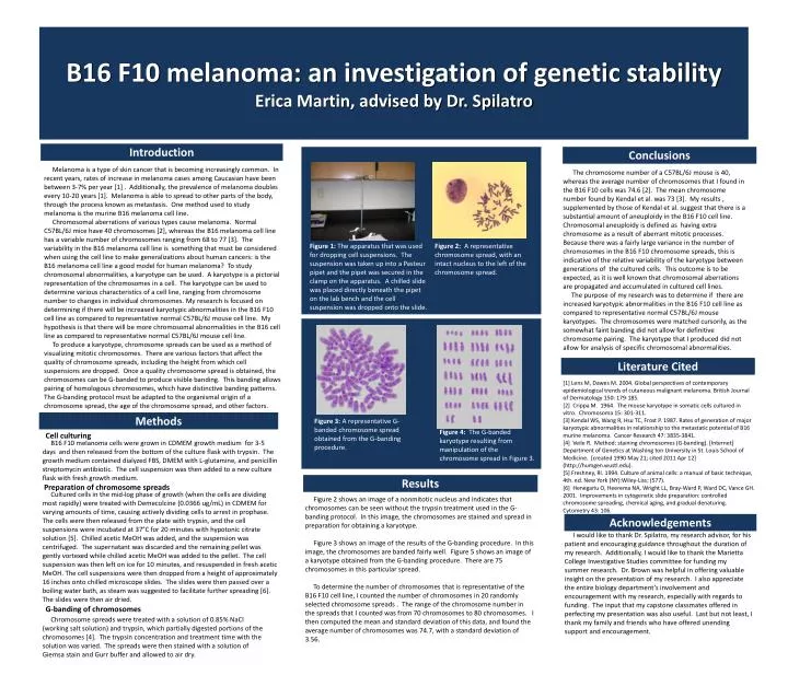
B16 F10 melanoma: an investigation of genetic stabilityErica Martin, advised by Dr. Spilatro Introduction Conclusions Melanoma is a type of skin cancer that is becoming increasingly common. In recent years, rates of increase in melanoma cases among Caucasian have been between 3-7% per year [1] . Additionally, the prevalence of melanoma doubles every 10-20 years [1]. Melanoma is able to spread to other parts of the body, through the process known as metastasis. One method used to study melanoma is the murine B16 melanoma cell line. Chromosomal aberrations of various types cause melanoma. Normal C57BL/6J mice have 40 chromosomes [2], whereas the B16 melanoma cell line has a variable number of chromosomes ranging from 68 to 77 [3]. The variability in the B16 melanoma cell line is something that must be considered when using the cell line to make generalizations about human cancers: is the B16 melanoma cell line a good model for human melanoma? To study chromosomal abnormalities, a karyotype can be used. A karyotype is a pictorial representation of the chromosomes in a cell. The karyotype can be used to determine various characteristics of a cell line, ranging from chromosome number to changes in individual chromosomes. My research is focused on determining if there will be increased karyotypic abnormalities in the B16 F10 cell line as compared to representative normal C57BL/6J mouse cell line. My hypothesis is that there will be more chromosomal abnormalities in the B16 cell line as compared to representative normal C57BL/6J mouse cell line. To produce a karyotype, chromosome spreads can be used as a method of visualizing mitotic chromosomes. There are various factors that affect the quality of chromosome spreads, including the height from which cell suspensions are dropped. Once a quality chromosome spread is obtained, the chromosomes can be G-banded to produce visible banding. This banding allows pairing of homologous chromosomes, which have distinctive banding patterns. The G-banding protocol must be adapted to the organismal origin of a chromosome spread, the age of the chromosome spread, and other factors. The chromosome number of a C57BL/6J mouse is 40, whereas the average number of chromosomes that I found in the B16 F10 cells was 74.6 [2]. The mean chromosome number found by Kendal et al. was 73 [3]. My results , supplemented by those of Kendal et al. suggest that there is a substantial amount of aneuploidy in the B16 F10 cell line. Chromosomal aneuploidy is defined as having extra chromosome as a result of aberrant mitotic processes. Because there was a fairly large variance in the number of chromosomes in the B16 F10 chromosome spreads, this is indicative of the relative variability of the karyotype between generations of the cultured cells. This outcome is to be expected, as it is well known that chromosomal aberrations are propagated and accumulated in cultured cell lines. The purpose of my research was to determine if there are increased karyotypic abnormalities in the B16 F10 cell line as compared to representative normal C57BL/6J mouse karyotypes. The chromosomes were matched cursorily, as the somewhat faint banding did not allow for definitive chromosome pairing. The karyotype that I produced did not allow for analysis of specific chromosomal abnormalities. Figure 1: The apparatus that was used for dropping cell suspensions. The suspension was taken up into a Pasteur pipet and the pipet was secured in the clamp on the apparatus. A chilled slide was placed directly beneath the pipet on the lab bench and the cell suspension was dropped onto the slide. Figure 2: A representative chromosome spread, with an intact nucleus to the left of the chromosome spread. Literature Cited [1] Lens M, Dawes M. 2004. Global perspectives of contemporary epidemiological trends of cutaneous malignant melanoma. British Journal of Dermatology 150: 179-185. [2] Crippa M. 1964. The mouse karyotype in somatic cells cultured in vitro. Chromosoma 15: 301-311. [3] Kendal WS, Wang R, Hsu TC, Frost P. 1987. Rates of generation of major karyotypic abnormalities in relationship to the metastatic potential of B16 murine melanoma. Cancer Research 47: 3835-3841. [4] Veile R. Method: staining chromosomes (G-banding). [Internet] Department of Genetics at Washing ton University in St. Louis School of Medicine. [created 1990 May 21; cited 2011 Apr 12] (http://humgen.wustl.edu). [5] Freshney, RI. 1994. Culture of animal cells: a manual of basic technique, 4th. ed. New York (NY):Wiley-Liss; (577). [6] Henegariu O, Heerema NA, Wright LL, Bray-Ward P, Ward DC, Vance GH. 2001. Improvements in cytogenetic slide preparation: controlled chromosome spreading, chemical aging, and gradual denaturing. Cytometry 43: 106. Methods Figure 3: A representative G-banded chromosome spread obtained from the G-banding procedure. Figure 4: The G-banded karyotype resulting from manipulation of the chromosome spread in Figure 3. Cell culturing B16 F10 melanoma cells were grown in CDMEM growth medium for 3-5 days and then released from the bottom of the culture flask with trypsin. The growth medium contained dialyzed FBS, DMEM with L-glutamine, and penicillin streptomycin antibiotic. The cell suspension was then added to a new culture flask with fresh growth medium. Results Preparation of chromosome spreads Cultured cells in the mid-log phase of growth (when the cells are dividing most rapidly) were treated with Demecolcine (0.0366 ug/mL) in CDMEM for varying amounts of time, causing actively dividing cells to arrest in prophase. The cells were then released from the plate with trypsin, and the cell suspensions were incubated at 37°C for 20 minutes with hypotonic citrate solution [5]. Chilled acetic MeOH was added, and the suspension was centrifuged. The supernatant was discarded and the remaining pellet was gently vortexed while chilled acetic MeOH was added to the pellet. The cell suspension was then left on ice for 10 minutes, and resuspended in fresh acetic MeOH. The cell suspensions were then dropped from a height of approximately 16 inches onto chilled microscope slides. The slides were then passed over a boiling water bath, as steam was suggested to facilitate further spreading [6]. The slides were then air dried. Figure 2 shows an image of a nonmitotic nucleus and indicates that chromosomes can be seen without the trypsin treatment used in the G-banding protocol. In this image, the chromosomes are stained and spread in preparation for obtaining a karyotype. Figure 3 shows an image of the results of the G-banding procedure. In this image, the chromosomes are banded fairly well. Figure 5 shows an image of a karyotype obtained from the G-banding procedure. There are 75 chromosomes in this particular spread. To determine the number of chromosomes that is representative of the B16 F10 cell line, I counted the number of chromosomes in 20 randomly selected chromosome spreads . The range of the chromosome number in the spreads that I counted was from 70 chromosomes to 80 chromosomes. I then computed the mean and standard deviation of this data, and found the average number of chromosomes was 74.7, with a standard deviation of 3.56. Acknowledgements I would like to thank Dr. Spilatro, my research advisor, for his patient and encouraging guidance throughout the duration of my research. Additionally, I would like to thank the Marietta College Investigative Studies committee for funding my summer research. Dr. Brown was helpful in offering valuable insight on the presentation of my research. I also appreciate the entire biology department’s involvement and encouragement with my research, especially with regards to funding. The input that my capstone classmates offered in perfecting my presentation was also useful. Last but not least, I thank my family and friends who have offered unending support and encouragement. G-banding of chromosomes Chromosome spreads were treated with a solution of 0.85% NaCl (working salt solution) and trypsin, which partially digested portions of the chromosomes [4]. The trypsin concentration and treatment time with the solution was varied. The spreads were then stained with a solution of Giemsa stain and Gurr buffer and allowed to air dry.