Download
1 / 59
590 likes | 730 Views
Sensory Receptors 8 th ed 50.1 to 50.4 7 th ed 49.1 to 49.4. Physiological basis for all animal activity: processing sensory information and generating motor output in response to that information . This is a continuous cycle

E N D
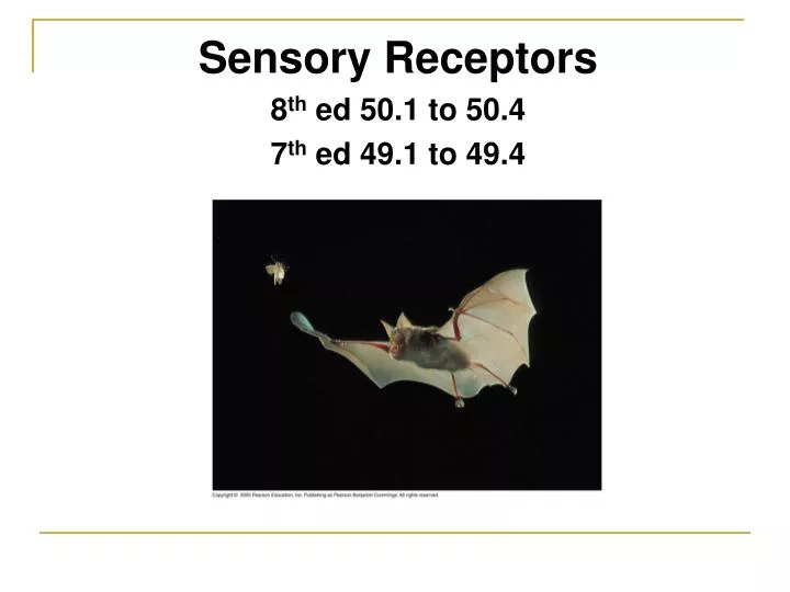
Sensory Receptors 8th ed 50.1 to 50.4 7th ed 49.1 to 49.4
Physiological basis for all animal activity: processing sensory information and generating motor output in response to that information
This is a continuous cycle • Sensory information can be from external or internal environment.
Sensory pathway: • Sensory reception • Transduction • Transmission • Perception • Amplification and adaptation
Sensory pathway: • Sensory reception • Transduction • Transmission • Perception • Amplification and adaptation
Sensory reception • 1st step of sensory pathway • Sensory receptors: Specialized neurons or epithelial cells; Single cells or a collection of cells in organs • Very sensitive
Sensory pathway: • Sensory reception • Transduction • Transmission • Perception • Amplification and adaptation
Sensory transduction: • Conversion of physical, chemical and other stimuli to change in membrane potential Receptor potential: • change in membrane potential itself
Sensory pathway: • Sensory reception • Transduction • Transmission • Perception • Amplification and adaptation
Transmission: • Sensory information is transmitted through the nervous system as nerve impulses or action potential to the Central Nervous System (CNS). • Some axons can extend directly into the CNS and some form synapses with dendrites of other neurons • Sensory neurons spontaneously generate action potential without stimulus at a low rate
Magnitude of receptor potential controls the rate at which action potentials are generated (larger receptor potential results in more frequent action potentials) Weak muscle stretch Strong muscle stretch Muscle Dendrites Receptor potential –50 –50 –70 –70 Stretch receptor Membrane potential (mV) Action potentials 0 0 Axon –70 –70 0 2 0 2 1 3 4 5 6 7 1 3 4 5 6 7 Time (sec) Time (sec)
Sensory pathway: • Sensory reception • Transduction • Transmission • Perception • Amplification and adaptation
Perception: • Action potential reach the brain via sensory neurons, generating perception of a stimulus • All action potentials have the same property, what makes the perceptions different are the part of the brain they link to.
Sensory pathway: • Sensory reception • Transduction • Transmission • Perception • Amplification and adaptation
Amplification and adaptation: • Strengthening of stimulus energy during transduction (involves second messengers) • Continued adaptation: decrease in responsiveness upon prolonged stimulation
Types of sensory receptors: • Mechanoreceptors • Chemoreceptors • Electromagnetic receptors • Thermoreceptors • Pain receptors
Mechanoreceptors: • sense physical deformation caused by pressure, touch, stretch, motion, sound
Stretch receptors are mechanoreceptors are dendrites that spiral around small skeletal muscle fibers Weak muscle stretch Strong muscle stretch Muscle Dendrites Receptor potential –50 –50 –70 –70 Stretch receptor Membrane potential (mV) Action potentials 0 0 Axon –70 –70 0 2 0 2 1 3 4 5 6 7 1 3 4 5 6 7 Time (sec) Time (sec) in the axon of the stretch receptor. A stronger stretch produces a larger receptor potential and higher frequency of action potentials. muscles and dendrites stretch, producing a receptor potential in the stretch receptor. The receptor potential triggers action potentials Crayfish stretch receptors have dendrites embedded in abdominal muscles. When the abdomen bends,
Hair Light touch Strong pressure • Touch receptors (light and deep touch) are embedded in connective tissue Epidermis Dermis Hypodermis Connective tissue Nerve
LE 49-4 Chemoreceptors: • General receptors: respond to total solute concentrations • Specific receptors: respond to concentrations of specific molecules
Eye Infrared receptor Electromagnetic receptors: • Detect various forms of electromagnetic energy like visible light, electricity, magnetism • Snakes can have very sensitive infrared receptors – detect body heat of prey • Animals can use earth’s magnetic field lines to orient themselves during migration (magnetite in body) – orientation mechanism This rattlesnake and other pit vipers have a pair of infrared receptors, one between each eye and nostril. The organs are sensitive enough to detect the infrared radiation emitted by a warm mouse a meter away. Some migrating animals, such as these beluga whales, apparently sense Earth’s magnetic field and use the information, along with other cues, for orientation.
Cold Hair Heat Thermoreceptors: • Detect heat and cold • Located in skin and anterior hypothalamus • Mammals have many thermoreceptors each for a specific temperature range Connective tissue Nerve
Capsaicin triggers the same thermoreceptors as high temperature • Menthol triggers the same receptors as cold (<28oC)
Hair Pain Pain receptors (nociceptors): • Stimulated by things that are harmful – high temperature, high pressure, noxious chemicals, inflammations • Defensive function Connective tissue Nerve
Sensing gravity and sound in invertebrates: • Hair of different stiffness and length vibrate at different frequencies and pick up sound waves and vibrations
Ciliated receptor cells • Statocyts: • organ with ciliated receptor cells surrounding a chamber containing statoliths in invertebrates – sense gravity Cilia Statolith Sensory nerve fibers
Tympanic membrane stretched over their “ear” help sense vibrations Tympanic membrane 1 mm
Sensing gravity and sound in humans: • Human ear: sensory organ for hearing and equilibrium • Our organ for hearing “hair cells” are mechanoreceptors because they respond to vibrations • Moving air pressure is converted to fluid pressure
Ear structure: • Outer ear: pinna, auditory canal, tympanic membrane (separates outer and middle ear)
Middle ear: three small bones malleus (hammer), incus (anvil) and stapes (stirrup) transmit vibrations from tympanic membrane to the oval window. • Eustachian tube connects middle ear to the pharynx and equalizes pressure • Inner ear: consists of fluid filled chambers including semicircular canals (equilibrium) and cochlea (hearing)
Moving air travels through the air canal and causes the tympanic membrane to vibrate • Three bones of the middle ear transmit the vibrations to the oval window, a membrane on the cochlear surface • That causes pressure waves in the fluid inside the cochlea
The cochlea: • Upper vestibular canal and inner tympanic canal filled with perilymph; middle cochlear duct filled with endolymph
Organ of Corti: • Floor of the cochlear duct is the basilar membrane • Organ of corti is located on the basilar membrane, with hair cells which has hair projecting into the cochlear duct. • Many of the hairs are attached to the overhanging tectorial membrane. • Sound waves cause the basilar membrane to vibrate. This results in displacement and bending of the hair cells within the bundle. • This activates the mechanoreceptors, changes the hair cell membrane potential (sensory transduction) which generates action potential in the sensory neuron.
Cochlea Stapes Axons of sensory neurons Vestibular canal Perilymph Oval window Apex Base Basilar membrane Tympanic canal Round window
Equilibrium: • In the vestibule behind the oval window are urticle, saccule and three semicircular canals
Three semicircular canals arranged in three spatial planes detect angular movements of the head • The hair cells form a cluster. They have a gelatinous capula. • Fluid in the semicircular canals pushes against the capula deflecting the hairs, stimulates the neurons Semicircular canals Ampulla Flow of endolymph Flow of endolymph Vestibular nerve Cupula Hairs Hair cell Vestibule Nerve fibers Utricle Body movement Saccule
Utricle (oriented horizontally), saccule (oriented vertically) tell the brian which way is up and the position of the body and acceleration • Sheet of hair cells project into a gelatinous capula embedded with otoliths (ear stones). Movement of the head causes otoliths to in different directions against the hair protruding from the hair cells. This movement is detected by the sensory neurons • Dizziness: false sensation of angular motion http://www.dizziness-and-balance.com/disorders/bppv/otoliths.html
Hearing and equilibrium in fish: • Vibrations in the water – conducted by skeleton to inner ear canals, move otoliths which stimulate hair cells • Swim bladder: air filled, responds to sound • Lateral line sense organ
Lateral line Lateral line sense organ: • Water flows through the system • Bends hair cells; generates receptor potential • Nerve carries action potential to the brain • Helps them sense water currents, moving objects, low frequency sounds Lateral line canal Scale Opening of lateral line canal Neuromast Epidermis Lateral nerve Segmental muscles of body wall Cupula Sensory hairs Supporting cell Hair cell Nerve fiber
Light Vision Photoreceptors • Planarians: Ocelli or eye spots in the head region • Light stimulates photoreceptors • Brain compares rate of action potential coming form the two ocelli • Brain directs the body to turn until sensation form both ocelli are equal and minimal • Animal can move to shade, under a rock away from predators Light shining from the front is detected Photoreceptor Nerve to brain Visual pigment Screening pigment Ocellus Light shining from behind is blocked by the screening pigment
Compound eyes: • Very good at detecting movement • Very good at detecting flickering light (6 times faster than human eye) • Some bees can see in the ultraviolet range of light
Several thousand omatidia (facets) in every eye • Cornea and crystalline cone form the lens which focuses light on the rhabdom • Light stimulates the photoreceptors to generate receptor potential which generates action potential Cornea Lens Crystalline cone Rhabdom Photoreceptor Axons 2 mm Ommatidium
Vertebrate eye • Single lens system (very different from invertebrate single eyes)
Choroid Sclera Iris Cornea • Eyeball or globe consists of • Sclera: tough white outer connective tissue • Cornea: clear part of sclera in the front of the eye – lets light into the eye, acts as a fixed lens • Choroid: pigmented inner layer: forms iris (doughnut shaped) – can change size to regulate the amount of light coming in Optic nerve Pupil Central artery and vein of the retina Optic disk (blind spot)
Retina • Retina: innermost layer with neurons and photoreceptors • Aqueous humor: fluid that fills the anterior cavity (blockage of ducts increases pressure and causes glaucoma) • Vitreous humor: jellylike, fills the posterior chamber Fovea (center of visual field) Aqueous humor Vitreous humor
Ciliary body • Lens: clear disk of protein Suspensory ligament Lens