Download

1 / 93
950 likes | 1.36k Views
Recent advances in MRI Breast and Future. Dr.Rattehalli R Ramachandra Consultant Radiologist University Hospitals Coventry & Warwick NHS trust. Introduction. Timeline of Breast diagnosis Role of MRI Breast Recent advances Other modalities Conclusion. Breast cancer UK.

E N D
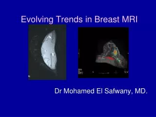
Recent advances in MRI Breast and Future Dr.Rattehalli R Ramachandra Consultant Radiologist University Hospitals Coventry & Warwick NHS trust
Introduction • Timeline of Breast diagnosis • Role of MRI Breast • Recent advances • Other modalities • Conclusion
Breast cancer UK • Commonest cancer in women • Accounts for 31% of all cancers in women • Life time risk for men 1 in 1014 • Life time risk for women 1 in 9 • Ref: Cancer research UK Feb 2009
Timeline of Breast Diagnosis • 1950’s – Breast Self Examination • 1960’s – BSE + Mammography • 1970’s – BSE + Mammography + Thermography+ Ultrasound • 1980’s – BSE + mammography + Better US • 1990’s – BSE + mammo + US + MRI • 2000’s – Digital mammo + US + MRI • 2010?? – Digital mammo + US + MRI + MR spectroscopy+Tomosynthesis + PEM + BSGI
Timeline of Breast Diagnosis • 1950’s – Breast Self Examination • 1960’s – BSE + Mammography • 1970’s – BSE + Mammography + Thermography+ Ultrasound • 1980’s – BSE + mammography + Better US • 1990’s – BSE + mammo + US + MRI • 2000’s – Digital mammo + US + MRI • 2010?? – Digital mammo + US + MRI + Tomosynthesis + PEM + BSGI
Sensitivity & SpecificityMammogram Vs Ultrasound Vs MRI Reference: HaithamElsamaloty et al . AJR 2009; 192:1142-1148, Increasing the accuracy of detection of Breast Cancer with 3-T MRI.
PPV of Mammography for Breast cancer For under 50 yrs ranges from 20% For age 50-69 yrs 60-80%
Sensitivity and Specificity of Annual MRI, Mammography, Ultrasound and 6 Monthly CBE in High Risk Women Cancer Imaging 2005; 5(1): 32-38
MR Vs Mammogram Examples • Netherlands study 1909 high risk patients 50 cancers 80% detected by MRI 33% detected by mammography
MR Vs Mammogram Examples • UK 649 high risk women 35 cancers MRI found 77% Mammography found 40%
MR Vs Mammogram Examples • Canada 236 Women at high risk 22 cancers MRI found 77% Mammo found 36%
MR Vs Mammogram Examples • Bonn 529 Women at high risk 43 cancers MRI found 91% Mammography found 33%
Breast Ultrasound • Not a screening test • Good for lumps • Good for clarification of abnormalities seen on mammography other than calcifications • Good for taking biopsies
DIGITAL MAMMOGRAPHY • DENSE BREASTS • WOMEN UNDER 50 • PREMENOPAUSAL WOMEN • EQUAL OR SLIGHTLY REDUCED RADIATION DOSE • Coventry is now fully digital • Digital Tomosynthesis reduces the recall rate in dense breasts
Indications • Staging newly diagnosed breast carcinoma ? • Lobular cancer staging • Unknown causes of axillary adenopathy • Neo adjuvant chemotherapy • Silicone implant rupture • Screening high risk patients • Radiation exposure at young age • Difficult mammogram/ultrasound/physical examination, Problem solving
COMICE Trial Results Between 2001 to 2007 1625 patients,817 with 807 without MRI Re operation with in 6 months was 18.8% with MRI & 19.3% without MRI Result: No significant benefit by addition of MRI to conventional Triple assessment Comparitive effeciveness of MRI in Breast cancer trial Reference: L.Turnbul,Symposium Mammographicum 2008.Lille, France 06/07/2008, Also Lancet 13/2/2010
Indications • Staging newly diagnosed breast carcinoma ? • Lobular cancer staging • Unknown causes of axillary adenopathy • Neo adjuvant chemotherapy • Silicone implant rupture • Screening high risk patients • Difficult mammogram/ultrasound/physical examination, Problem solving • Radiation exposure at young age
MRI in Invasive Lobular cancer MRI accurately assesses the size & extent of cancer Detects cancer on other side Can change treatment plan in up to 28% of cases NICE guideline
P W 2006 HISTORY • 55YRS OLD • P 3 R4 LUMP IN RIGHT BREAST • US BIOPSY B5b LOBULAR SINGLE LESION • MRI TO EXCLUDE ANY OTHER LESION • OTHERWISE SUITABLE FOR WLE
Indications • Staging newly diagnosed breast carcinoma ? • Lobular cancer staging • Unknown causes of axillary adenopathy • Neo adjuvant chemotherapy • Silicone implant rupture • Screening high risk patients • Difficult mammogram/ultrasound/physical examination, Problem solving • Radiation exposure at young age
Metastatic Nodes in Axilla With No Obvious Primary in Breast < 2% of patients present with palpable axillary nodes and negative mammogram and US MRI finds the primary in up to 60-75% of cases This should be confirmed by second look US or MR guided biopsy
Indications • Staging newly diagnosed breast carcinoma ? • Lobular cancer staging • Unknown causes of axillary adenopathy • Neo adjuvant chemotherapy • Silicone implant rupture • Screening high risk patients • Difficult mammogram/ultrasound/physical examination, Problem solving • Radiation exposure at young age
Silicon only image. Extra capsular silicon with fluid collection
Indications • Staging newly diagnosed breast carcinoma ? • Lobular cancer staging • Unknown causes of axillary adenopathy • Neo adjuvant chemotherapy • Silicone implant rupture • Screening high risk patients • Radiation exposure at young age • Difficult mammogram/ultrasound/physical examination, Problem solving
New ACS Guidelines for Annual MRI Screening in addition to Mammo(May, 2007) • Any woman who has greater than 20% lifetime risk of developing breast cancer (BRACAPRO, GAIL, BOADACEA) • BRCA mutation and untested relatives • Prior XRT (bet ages of 10-30)
NICE Guideline MRI annual surveillance • From 30-39 yrs: • To women at a 10 year risk >8% • From 40-49 yrs: • To women at 10 year risk of > 20% or • To women at a 10 year risk of > 12% where mammography has shown a dense breast pattern
Radiation exposure at young age • Hodgkin's disease treated with Mantle radiation • Risk of BC increases beginning about 7-8yrs after treatment peaking at about 15yrs post treatment • Younger age at treatment = Higher risk • Many unaware of risk • Begin intensive screening 6-7 yrs after treatment
Indications • Staging newly diagnosed breast carcinoma ? • Lobular cancer staging • Unknown causes of axillary adenopathy • Neo adjuvant chemotherapy • Silicone implant rupture • Screening high risk patients • Radiation exposure at young age • Difficult mammogram/ultrasound/physical examination, Problem solving
Problem solving Case 1
SH 60 yrs. Recalled from screening for possible ASD Right Breast
MDM Specimen X ray normal Breast tissue HP: No tumour in the specimen SNB positive Repeat US: Post operative changes only with lot of oedema and seroma. No tumour seen Decision: To do MRI to try and Identify the tumour