Download
1 / 67
680 likes | 842 Views
Blood and the Circulatory System. Functions of Blood. A “closed” system of fluid delivery Red blood cells never leave the blood vessels Immune cells must receive a signal in order to leave the blood vessels Fluids & gasses can only leave via the capillary network

E N D
Functions of Blood • A “closed” system of fluid delivery • Red blood cells never leave the blood vessels • Immune cells must receive a signal in order to leave the blood vessels • Fluids & gasses can only leave via the capillary network • Fluids & gasses are contained within the blood plasma • Transports carbohydrates, fats, amino acids, proteins, hormones, blood cells, electrolytes & water to every part of the body
Blood • Total blood volume 5L • Roughly 8-10% total body weight • Thicker viscosity than water (obviously…there are cells and proteins in it) • pH 7.35-7.45 (recall pH homeostasis) • Normally 38C • When you donate blood, 1 “unit” = 500 ml (10% total blood volume) • 2 component groups: • Formed elements (45% total volume) • Blood plasma (fluid portion)
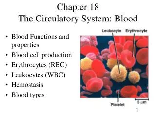
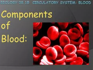
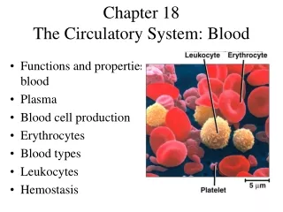
Parts of Blood • “formed elements” & blood plasma • Formed elements = cells of the blood • Red blood cells (erythrocytes) • Carry oxygen • Leukocytes (white cells) • Granulocytes (have vesicles within them that can be seen) • Neutrophils, eosinophils & basophils • Agranular leukocytes (do not have vesicles within them) • Monocytes/macrophages & lymphocytes (T- & B-cells) • Platelets (cell fragments that initiate blood clots) • Blood plasma = fluid portion of blood • Where the nutrients, electrolytes, water & dissolved gasses can be found
RBC’s & Platelets • Platelets: • Shortest lifespan ( 5-7 days) • Cell fragments (originate in bone marrow from “megakaryocyte”) • No nucleus • During blood vessel damage, platelets will “aggregate” and begin the clot formation • Red blood cells (RBC, erythrocytes): • 120 day lifespan • No nucleus, bi-concave shape • Shape increases surface area for gas exchange • Also permits cell flexibility • Hemoglobin: oxygen carrying molecule
Platelets & Blood Clots • Platelets can sense a damaged blood vessel • Will migrate and attempt to adhere (aggregate) at site of vessel damage • After the initial aggregation, platelets then draw in “fibrinogen” (large protein found in plasma) • Fibrinogen acts like a wicker basket to hold the clot together • Hemophilia: genetic defect in one of the many proteins involved in clot formation • Platelet proteins, fibrinogen etc.
Recent information into the way platelets form from a megakaryocyte indicate that the megakaryocyte “sticks out” a cellular appendage that gets “broken off” in the blood stream.
Hemoglobin • 4 subunit protein that carries oxygen & carbon dioxide (CO2) • Requires iron: iron is what actually holds onto the oxygen • Changes shape when bound to oxygen (why oxygen-rich blood looks bright red & “deoxygenated” blood looks brown/maroon) • Usually 250-300 billion hemoglobin molecules per RBC • Allows one RBC to capture 1 trillion O2 molecules
Anemia • Refers to either decreased red blood cell count OR decreased oxygen carrying capacity • Pernicious anemia = vitamin B12 deficiency = low RBC count • Autoimmune disease : reduced dietary B12 absorption = gradual & progressive loss of liver B12 stores (B12 required by bone marrow for RBC production) • Aplastic anemia = chemical destruction of red bone marrow = low RBC count • Microcytic anemia = defect in hemoglobin production (usually due to low iron stores) • Sickle-cell anemia = genetic defect in hemoglobin sequence • Regions of heme molecule cannot hold O2 and subsequently alter the shape of the RBC • Defective hemoglobin = altered RBC shape (often leads to inappropriate clot formation)
Granular leukocytes (granulocytes) • “Grains” = vesicles of digestive enzymes or chemicals • Recall peroxisomes & lysosomes • Like to eat things and blow stuff up • Neutrophils = most common • Eosinophils = primarily for parasitic defense • Endocytose parasites and fuse granules to the parasite to kill • Basophils = primarily for blood vessel “control” • Granules contain histamine • Not so much for allergies, more for local “vasodilation”…increases blood vessel size/diameter so that more blood can get to the site
Agranular Leukocytes • More focused on endocytosis (prefer to eat targets rather than blow them up) • Monocytes/macrophages: • Largest cells in the blood (called monocyte) • Often leave the blood and enter tissue (then called macrophage) • Lymphocytes: • Most common agranular lymphocyte • T-cells = acquired immune cells • Need to be “activated”, will attack a known antigen • B-cells = antibody-producing cells
Antibody • Antibody = protein made by “adaptive immune system” • Agranular leukocytes (T-cells & B-cells) comprise the “adaptive immune system) • Adaptive because they “learn” throughout childhood • When you are exposed to a new virus or bacteria, T-cells initiate the “learning” process • B-cells then “trained” to produce antibody • Antibody is how the adaptive immune system “recognizes” a target
Antibody • B-cells constantly produce antibody • During an infection, thymus stimulates specific B-cells to “clone” or increase in number • Those B-cells that produce the antibody that recognizes the invader • As the antibody is made and released into the blood, it will bind to its target • Once bound , will stimulate other immune cells (neutrophils, monocytes, Killer-T cells etc.) to kill or eat that target
Antibody • Antibodies are made by B-cells • Each B-cell produces 1 specific antibody • If your immune system can produce 1000 antibodies, you will have at least 1000 different B-cells • Antibody is a protein, works like an enzyme • Recognizes a specific part, structure etc. on a target • Adaptive immune system knows how to make antibodies to ANYTHING • Virus, bacteria, protein etc. • “anti-body” = against a foreign body
Antibody • Adaptive immune system “learns” during childhood • The more you expose the immune system to, the more it can recognize • After maturity, immune system relies on “revising” it’s “repertoire” rather than learning anything new • B-cells can be trained to “re-work” their current antibody to recognize a new invader • ONLY to a certain degree (there is a limit as to how much a B-cell can “revise” its antibody)
“Closed System” • Recall red blood cells will never leave the circulatory blood vessel system (they never leave the “tube”) • White cells (leukocytes…don’t confuse these with the sub-type lymphocytes) CAN leave the blood vessels • “diapedesis” or “extravasation” • Diapedesis = ameba-like cell movement • How immune system can leave the blood and enter a tissue to deal with pathogens
http://video.google.com/videoplay?docid=-142799799667345732&q=diapedesis&total=1&start=0&num=10&so=0&type=search&plindex=0http://video.google.com/videoplay?docid=-142799799667345732&q=diapedesis&total=1&start=0&num=10&so=0&type=search&plindex=0
Blood Plasma • Remember that blood is a form of “connective tissue” • Connective tissue has an extracellular matrix or “ground substance” (materials other than the cells themselves)…like cartilage and bone • Blood plasma is this extracellular matrix (not a solid…a liquid matrix) • Yellowish colored fluid (yellow from all the protein)
Blood Plasma • Within the plasma: • Glucose, amino acids, lipids (carried by a protein), water, electrolytes (various ions) • “Blood proteins” or “plasma proteins” • Almost all produced by the liver • Albumins (large carrier protein…like a semi) • Globulins (lipid carrier proteins) • Subtype called gamma globulins = antibodies (made by B-cells…a type of lymphocyte) • Fibrinogen (largest protein in blood…for blood clot formation…works after the platelets have “aggregated”) • The combination of these large proteins is what colors plasma yellow
Blood Plasma • Important facts: • Remember that the cells of the blood don’t carry the nutrients • Carbohydrates (glucose), amino acids & lipids (carried by a protein) are all “water soluble” & dissolved within the blood plasma • The movement of the cells through the blood vessel network “pushes” the plasma along • Where red blood cells cannot leave the blood vessel network, the plasma can readily “leak” out to the nearby cells in a capillary bed • To deliver these nutrients and gas to all the cells of your body (no cell is more than a few microns from a capillary)
Blood Plasma • Important facts: • Ingredients of blood plasma are important for osmotic control • It is the fluid that all your cells are “suspended” in • Plasma proteins are vital for controlling osmotic balance • Too many proteins/electrolytes = hypertonic = dehydrate the cells (draw out water to dilute the plasma) • Too few proteins = hypotonic = swell the cells (force water to push into the cells)
Blood Plasma Hyper- vs. Hypotonic blood plasma Normal blood plasma = isotonic (equal tonic pressure) Hypertonic = too much solutes (too many particles) in the plasma In essence, the cells of your body will have more water molecules/particles WITHIN them than the plasma Will result in water LEAVING the cells to dilute the plasma (because the solutes/particles in plasma cannot easily enter the cells)
Blood Plasma Hyper- vs. Hypotonic blood plasma Normal blood plasma = isotonic (equal tonic pressure) Hypotonic = too few particles in the plasma compared to what is in each cell Result is too much water outside the cell…water will then enter the cells to balance the concentrations So that there are will be an equal ratio water:particle in BOTH the plasma AND the cells
Blood Plasma Electrolytes (sodium/Na+, potassium/K+) are KEY players in blood plasma “tonicity” Have an electrical charge (cannot easily pass across a cell’s plasma membrane) Concentration in blood plasma AND within the cells of your body must be tightly regulated Electrolyte concentration in your blood is not only important for controlling the movement of water in/out of your cells, but also for the electrical gradient that each cell maintains Remember how muscle cells and neurons get excited?
Blood Plasma • Important facts: • Plasma brings nutrients and oxygen TO the tissues, and also carries away metabolic waste & CO2 FROM those tissues • The gasses (oxygen & CO2) have to dissolve in the fluid portion in order to transfer to the target tissue • The RBC’s do not directly transfer oxygen and pick up CO2 • Rely on a concentration gradient… • When they enter a region of low oxygen in the blood plasma (where the nearby cells are taking up oxygen at a high rate), the concentration of oxygen outside the RBC favors losing the oxygen into the plasma • This can ONLY occur in a capillary…only a capillary is “porous” enough to allow plasma to pass across
Blood Types • 4 blood types: A, B, AB & O • Defined by presence of specific antibody • Type A blood has antibodies against Type B blood (you have antibodies against the other type…no sense to have antibodies against your own blood type) • Type AB has NO antibodies (can receive blood from any donor… “universal recipient”) • Type O = BOTH antibodies (Type A and Type B antibodies) • “Universal donor”
Blood Types • Tested by mixing blood with each respective antibody (agglutination) • Type A blood with Type A antibody will clump… • You cannot give this blood to a patient who is Type B (because they have antibodies against Type A blood and it will clot) • Since Type AB has NO antibodies, these patients can receive blood from any donor (they will not clot or agglutinate the donated blood)
Blood Types • Positive & negative refers to “Rh factor” • An ANTIGEN (not antibody) (Rh = rhesus macaque) • Positive (+) = have Rh antigen/protein • Significant for fetal development: • If mother is Rh (-) (no antigen/protein), and fetus genetic disposition = Rh (+) • Possible if the father is Rh (+) • Risk is that mother will develop antibodies against the Rh factor/antigen/protein and mount an immune response against fetus • What normally occurs = mother’s immune system will attack red blood cells of the fetus
Blood Types • Negative mother & positive fetus: • As mother’s immune system attacks fetal blood, fetus can become anemic • “hemolytic disease of the newborn” or fetal anemia • Common cause of stillborn births • Can be managed: first diagnosed by parental blood typing • If there is a risk, mother is usually given “RhoGAM” injections prior to birth • A mixture of Rh antibodies that “blinds” the mothers immune system from seeing the fetal Rh factor/protein so that her immune system does not mount a response
Blood Types • Type A positive: • “Plasma has antibodies against Type B blood, and is positive for the Rh antigen” • Type A negative: • “Plasma has antibodies against Type B blood, and does NOT have Rh antigen”
Circulatory System • Need a system in order to propel blood to every region of the body • Circulatory system = heart & blood vessels • Recall: blood vessel network is a “closed system” • Pores in the blood vessels are not large enough to permit cells to easily pass • Immune cells have to “squish” their way through the pores • Red blood cells never leave the vessel tube network
Heart • 4 – chamber “double pump” muscle • Pumps 5 L/min (2.5+ million liters year) • At roughly 60-70 bpm, heart pumps almost 50 million times / year • An RBC takes 1 minute to travel from the heart to your finger or toe and back to the heart! • Roughly size of your fist • Contained within pericardium • Sac of connective tissue that forms a bag around the heart
Cardiac muscle • Most of your heart is made of cardiac muscle • Striated (like skeletal muscle in contractile protein arrangement) • Each cell is much shorter, and usually more thick as well • Each cell joined by an intercalated disc • An area of cell-cell adhesion, as well as a gap junction complex to permit 1 cell to stimulate the next
Cardiac muscle • Cardiac muscle cells have less developed sarcoplasmic reticulum • Less ability to store calcium • Reliant upon plasma calcium rather than “stored” calcium (contrast to skeletal muscle cells) • Damage is repaired by fibrosis (scar tissue) ONLY • Reason why heart attack patients have such a hard time recovering…they’ve lost heart contractility and will never regain/repair it • In contrast, if you damage skeletal muscle, some of it will repair itself (“satellite cells” within skeletal muscle can form new skeletal muscle cells)
Cardiac muscle • NO neural stimulus for contraction • Has “pacemaker cells” that set off rhythmic depolarizations (electrical pulses) to trigger your heartbeat • The autonomic nervous system feeds into these pacemaker cells to increase or decrease your heart rate, but these cells operate without neural control to make sure you heart ALWAYS beats • Known as “autorhythmic” because your heart does not need your brain to tell it to beat • Note: these are neurons, just not in your BRAIN or SPINAL CORD • Pacemaker cells in your heart are specialized neurons that have a “cycle”…why they’re called pacemakers
Heart • 4 chambers & valves • Atria are the initial “receptacles” of the heart • Interatrial septum = thin, muscular membrane separating left & right atria • Ventricles = much more muscular (the actual “pump” regions • Intraventricular septum = thick muscular membrane separating left & right ventricles • Valves maintain one-way flow of blood through heart • Atrioventricular valves separate atria from ventricles • “semilunar” valves separate ventricles from the aorta & pulmonary vein
Heart Blood circuit: Venous blood enters heart via right atrium Atrium contracts & sends blood into right ventricle Ventricle contracts and sends blood into pulmonary artery into lungs Note: this artery contains oxygen-poor blood Left atrium receives oxygen-rich blood from lungs via pulmonary vein Note: this vein contains oxygen-enriched blood Atrium contracts & sends blood into left ventricle Ventricle contracts & sends blood into the aorta (largest artery in the body)
Heart Blood circuit notes: BOTH atria contract at the same time BOTH ventricles contract at the same time Images in the books are misleading Right ventricle often appears the same size as the left ventricle… In fact, right ventricle is often “thinner” or less muscular than the left ventricle Whereas the right ventricle only needs to pump blood to the lungs, the left ventricle must pump blood throughout the body (hence, thickness of the left ventricle wall is greater)
Why does the inside of your heart look so “rough”? Imagine if it were perfectly smooth…whenever it contracted, the walls would “stick together” like a wet plastic bag…the roughness prevents this from happening (imagine placing a layer of sponges inside that wet plastic bag).
Heart The pump cycle is self-perpetuating Recall the “reverberating neural circuit” Neurons within the heart depolarize in a cycle (60-100 times per minute) These depolarizations begin in the sinoatrial node (SA note…the pacemaker “region” of the heart) Depolarization spreads throughout BOTH atria Wave is carried/focused onto the atrioventricular node (AV node) Wave passes from AV node down through the intraventricular septum Depolarizations then fan outward from the bottom (apex) of the heart throughout the ventricles
Heart Conduction Heart sounds: 2 distinct sounds: “lub=dub” = valves slapping shut Lub = Atrioventricular valves closing (ventricles contracting, SYSTOLE) Dub = aortic & pulmonary valves closing (ventricles relaxing, DIASTOLE) Blood pressure = “120/80” refers to systole / diastole pressure 120 mm/Hg systolic pressure (ventricles contract and produce 120 mm/Hg blood pressure “spike”) 80 mm/Hg diastolic pressure (when ventricles are relaxed)
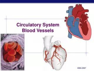
Blood vessels • Tubular network for blood flow • 3 layers to every blood vessel • Tunica externa • Layer that you can see when you cut down to expose the vessel • Tunica media • Smooth muscle layer • In arteries, tunica media layer contains large density of elastic fibers & is VERY thick • Tunica interna (endothelium) • Specialized connective tissue that permits fluid and gas exchange, but keeps the cells of the blood restricted within the blood vessel
Blood vessels Since the blood vessels form a “closed loop” system, the pathway of blood is predictable Bear in mind that an artery carries blood AWAY from the heart, veins carry blood TO the heart (these terms have NOTHING to do with the amount of oxygen in the blood).
Blood vessels Major arteries carry blood to a region of the body Smaller arteries branch off within this region Smaller arterioles then branch off to diversify the coverage Blood gasses, nutrients & plasma can only leave the vessel system via the capillary network Capillary bed = network of capillaries that services a particular area of tissue Can often be “mapped” Mapping allows you to remove a particular “capillary bed” surgically (ie. if you need to remove a carcinomic growth)