Download
1 / 1
10 likes | 110 Views
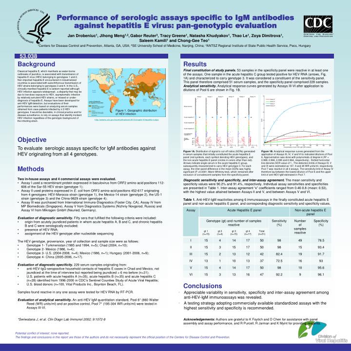
Figure 1. Geographic distribution of HEV infection. Performance of serologic assays specific to IgM antibodies against hepatitis E virus: pan-genotypic evaluation. Jan Drobeniuc 1 , Jihong Meng 1,2 , Gabor Reuter 3 , Tracy Greene 1 , Natasha Khudyakov 1 , Thao Le 1 , Zoya Dimitrova 1 ,

E N D
Figure 1. Geographic distribution of HEV infection Performance of serologic assays specific to IgM antibodies against hepatitis E virus: pan-genotypic evaluation Jan Drobeniuc1, Jihong Meng1,2,Gabor Reuter3, Tracy Greene1, Natasha Khudyakov1, Thao Le1, Zoya Dimitrova1, Saleem Kamili1 and Chong-Gee Teo1 1Centers for Disease Control and Prevention, Atlanta, GA, USA; 2SE University School of Medicine, Nanjing, China; 3ANTSZ Regional Institute of State Public Health Service, Pecs, Hungary 53.038 Background Results Final constitution of study panels. 53 samples in the specificity panel were reactive in at least one of the assays. One sample in the acute hepatitis C group tested positive for HEV RNA (arrows, Fig. 1A) and characterized to carry genotype 3. It was considered a constituent of the sensitivity panel. This panel therefore comprised 51 serum samples, and the specificity panel comprised 228 samples. Analytical sensitivity.Analytical response curves generated by Assays III-VI after application to dilutions of Pool 6 are shown in Fig. 1B. Classical hepatitis E, which manifests as water-borne outbreaks of jaundice, is associated with transmission of hepatitis E virus (HEV) belonging to genotypes 1 and 2. Non-imported hepatitis E encountered in industrialized countries is associated with autochthonous transmission of HEV strains belonging to genotypes 3 and 4. In the U.S., clinically manifest hepatitis E is seldom reported although HEV infection appears widespread , a disparity that may be due to low-dose exposure to HEV, asymptomatic infection by relatively avirulent HEV strains, or missed or under-diagnosis of hepatitis E. Assays have been developed for anti-HEV IgM detection, but evaluations of their performances were based on analyzing serum samples obtained from case-patients infected by ≤ 2 HEV genotypes. It would be desirable, in clinical practice and for disease surveillance, to rely on assays that identify incident HEV infection regardless of the genotypic background of the infecting strain. http://wwwnc.cdc.gov/travel/yellowbook/2010/chapter-5/hepatitis-e.aspx Objective To evaluate serologic assays specific for IgM antibodies against HEV originating from all 4 genotypes. Figure 1A. Distribution of signal to cut-off ratios (SCRs) generated in serum samples that initially constituted the acute hepatitis E panel (red symbols, each symbol denoting HEV genotypes), and the non-acute hepatitis E panel (circles in colors other than red). Arrows indicate single serum in the acute hepatitis C group, subsequently characterized to carry HEV genotype 3. For each assay, the inter-panel difference in the mean SCRs was highly significant (P <0.0001; Mann-Whitney test), which remained after exclusion of convalescent samples from the specificity panel. Figure 1B. Analytical response curves generated from the application of Assays III, IV, V and VI to indicated dilutions of Pool 6. Approximation was done with polynomials of degree 4 (R2 = 0.989, 0.966, 0.995 and 0.964, respectively). Dotted horizontal line denotes SCR value of 1.. The detection limits of Assays III, V and VI were estimated as 137, 9 and 49 WR units/ml, respectively. Pool 7 was reactive in all 4 assays. The sensitivity limit of Assay IV therefore lay between the lowest dilution of Pool 6 and the upper limit of anti-HEV IgM estimated in Pool 7. Methods • Two in-house assays and 4 commercial assays were evaluated. • Assay I used a recombinant protein expressed in baculovirus from ORF2 amino-acid positions 112-606 of the Sar-55 HEV strain (genotype 1); • Assay II used proteins expressed in E. coli from ORF2 amino-acid positions 452-617 originating from 4 genotypes: HEV-Morocco strain (genotype 1), the Mexico-14 strain (genotype 2), the US-1 strain (genotype 3) and the China-9829 strain (genotype 4); • Assay III was purchased from International Immuno-Diagnostics (Foster City, CA), Assay IV from MP Biomedicals (Singapore), Assay V from Diagnostics Systems (Nizhniy Novgorod, Russia) and Assay VI from Mikrogen GmbH (Neuried, Germany). • Evaluation of diagnostic sensitivity. Fifty sera that fulfilled the following criteria were included: • origin from acutely jaundiced patients in whom acute hepatitis A, B and C, and chronic hepatitis B and C were serologically excluded; • presence of HEV RNA; • assignment of the HEV genotype after nucleotide sequencing • The HEV genotype, provenance, year of collection and sample size were as follows: • Genotype 1- Turkmenistan (1985 and 1994, n=5); Chad (2004, n=10); • Genotype 2- Mexico (1986, n=4); • Genotype 3- U.S. (2004-2008, n=4); Mexico (1986, n=1); Hungary (2001-2006, n=9); • Genotype 4- China (2005-2006, n=17). • Evaluation of diagnostic specificity. 229 serum samples originating from: • anti-HEV-IgG-seropositive household contacts of hepatitis E cases in Chad and Mexico, not jaundiced at the time of interview but reported being jaundiced > 6 mo before (n=31); • U.S. patients with acute hepatitis A (n=35), acute hepatitis B (n=35) and acute hepatitis C (n=28) identified from 1996-2006 in CDC’s Sentinel Counties Study of Acute Viral Hepatitis; • U.S. blood donors (n=100, Vital Products Inc., Boynton Beach, FL). • Samples found reactive in any one assay were tested for HEV RNA by RT-PCR. • Evaluation of analytical sensitivity.An anti-HEV IgM quantitation standard, Pool 6* (860 Walter Reed (WR) units/ml) and an positive control, Pool 7* (195-304 WR units/ml) were tested in Assays III-VI. • *Seriwatana J, et al. ClinDiagn Lab Immunol 2002; 9:1072-8 Diagnostic sensitivity and specificity, and inter-assay agreement. The mean sensitivity and specificity values were 90.2% and 91.4%, respectively. Individual assay sensitivities and specificities are presented in Table 1. Inter-assay agreement “κ” coefficients ranged from 0.46-0.8 (mean: 0.53), with the highest value attained between Assays II and V, and between Assays V and VI. Table 1. Anti-HEV-IgM reactivities among 6 immunoassays in the finally constituted acute hepatitis E panel and non-acute hepatitis E panel, and corresponding diagnostic sensitivity and specificity values. Conclusions • Appreciable variability in sensitivity, specificity and inter-assay agreement among anti-HEV-IgM immunoassays was revealed. • A testing strategy adopting commercially available standardized assays with the highest sensitivity and specificity is recommended. Acknowledgements: Authors are grateful to K Foytich and D Chen for assistance with panel assembly and assay performance, and R Purcell, R Jarman and K Myint for providing reagents. Potential conflict of interest: none reported.