Download
1 / 19
200 likes | 351 Views
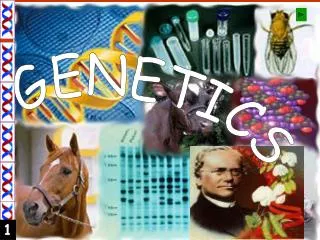
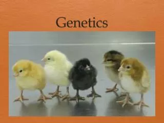
Genetics. Genetics is the study of genes Genes; sequences of DNA. Genes are packaged together as chromosomes and are passed from parent to offspring.

E N D
Genetics • Genetics is the study of genes • Genes; sequences of DNA. • Genes are packaged together as chromosomes and are passed from parent to offspring. • It is our genes that determine who we are and how we function at the most basic cellular level. Sometimes mistakes (mutations) cause significant disability or death, or benefits. • Genes; 25,000 genes.
Chromosomes • Chromosomes are made up of molecules of DNA, complexed with proteins called histones. • Chromosomes together carry the genetic blueprint of an individual. • All human somatic (body) cells contain 23 pairs of chromosomes, one pair from each parent, for a total of 46 chromosomes. Each human sex cell, an egg or a sperm, contains 23 unpaired chromosomes.
Gene Activation Although each somatic cell contains the same 23 pairs of chromosomes, only certain genes are activated in any given cell; therefore, only certain proteins or enzymes are produced by that cell. Which genes are activated in which cell is determined during embryologic development and throughout life by circulating growth factors, hormones, and chemical produced by a given cell and its neighboring cells.
Cellular Reproduction • All cells reproduce during embryonic development, which allows for growth of the embryo and differentiation (specialization) of the cells making up tissues and organs. • After birth and throughout adulthood, many cells continue to reproduce. • Cells that reproduce throughout a lifetime include cells of the bone marrow, skin, and digestive tract. • Liver and kidney cells reproduce when replacement of lost or destroyed cells is required. Special cells, called stem cells, are capable of reproducing indefinitely. • Other cells, including nerve, skeletal muscle, and cardiac muscle cells, do not reproduce significantly after the first few months following birth.
The Cell Cycle The cell cycle refers to a sequence of stages a cell goes through during its lifetime . During embryogenesis, all cells go through all stages, as do adult cells that continue to reproduce. The rate at which a cell goes through its cell cycle depends on the given cell and the growth factors, hormones, and chemicals to which it is exposed. Cells that do not continue to reproduce after embryogenesis remain in a resting stage and do not cycle through the other stages. The cell cycle is divided into two parts: interphase and mitosis.
Interphase • When not actively dividing, a cell is said to be in interphase. Mitosis • Mitosis is the stage of cell division. Mitosis is a shorter event than interphase; it lasts approximately 1 hour. During mitosis, the cell that has duplicated during interphasesplits into two daughter cells that each contain the 23 pairs of chromosomes. • Mitosis consists of the substages of prophase, metaphase, anaphase, and telophase
Meiosis • Meiosis is the process during which germ cells of the ovary (primary oocytes) or testicle (primary spermatocytes) give rise to mature eggs or sperm . • Meiosis involves DNA replication in the germ cell, followed by two cell divisions rather than one, which results in four daughter cells, each with 23 (unpaired) chromosomes. • In males, all four daughter cells are viable and continue to differentiate into mature sperm. • In females, only one viable daughter cell (egg) is formed; the other three cells become nonfunctional polar bodies. • During fertilization, genetic information contained in the 23 chromosomes of the egg joins with genetic information contained in the 23 chromosomes of the sperm. This results in an embryo with 46 total chromosomes (two pairs of 23).
An interesting phenomenon occurs during DNA replication in the first meiotic stage. At this time, pieces of DNA may shift between the matched chromosome pairs, in a process called crossing-over. Crossing-over increases the genetic variability of offspring, and is one reason why siblings within a family may vary considerably in genotype and phenotype.
Genotype and Phenotype • Precise genetic information carried in the chromosomes of the offspring is termed the genotype. Physical representation of genetic information (tall or short, dark or light) is called the phenotype. Genetic Testing (cytogenetics) • Genetic testing, called cytogenetics, involves looking at the overall structure and number of the chromosomes. Genetic testing can be performed on any cell of the body, but in children and adults it is usually done by withdrawing white blood cells in a venous blood sample. For prenatal testing, fetal cells may be gathered during the processes of amniocentesis, or during chorionic villi sampling.
Amniocentesis • Amniocentesis is performed by inserting a needle through the abdominal wall of a pregnant woman into the amniotic sac that surrounds the fetus. Amniotic fluid, into which fetal cells have been shed, is withdrawn. Chromosomes present in the fluid sample are then cultured and fixed, and their number and shape are analyzed for genetic integrity. This test is usually done at approximately 16 weeks' gestation and results are available in approximately 2 weeks.
Chorionic Villi Sampling • Chorionic villi sampling involves gathering cells of the chorion , the outer border of the fetal membranes. The cells are gathered by placing a needle through the woman's lower abdomen or cervix between 8 and 12 weeks of pregnancy. The cells do not need to be cultured, so the chromosomal analysis is available in approximately 1 to 2 days..
Pathophysiologic Concepts Mutation: • A mutation is an error in the DNA sequence. Mutations can occur spontaneously, or after the exposure of a cell to radiation, certain chemicals, or various viral agents. • Most mutations will be identified and repaired by enzymes working in the cell. • If a mutation is not identified or repaired, or if the cell does not undergo programmed death, that mutation will be passed on in all subsequent cell divisions. • Mutations may result in a cell becoming cancerous. Mutations in the gametes (the egg or sperm) may lead to congenital defects in an offspring
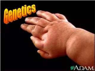
congenital Defects • Also called birth defects, include genotypic and phenotypic errors occurring during embryogenesis and fetal development. • Some congenital defects, such as cleft palate and limb abnormalities, may be apparent at birth, whereas other congenital defects, such as an abnormal or absent kidney and certain types of heart disease, may not be recognized immediately. • Congenital defects may result from genetic mistakes made during meiosis of the sperm or egg, or from environmental insults experienced by the fetus during gestation.
Chromosomal Breaks • During mitosis and meiosis, pieces of chromosomes may break off, be added inappropriately to other chromosomes, or be deleted entirely. If deletions or additions occur during meiosis in the egg or sperm, a congenital defect or death of the embryo may result. • If deletions or additions of chromosomes occur during mitosis, the affected cell line will usually die out.
Errors in Chromosome Number • Any change from the normal human chromosome number of 46 chromosomes is called aneuploidy. • An aneuploidy in which there are only 45 chromosomes is called a monosomy. • An aneuploidy in which there are 47 chromosomes is called a trisomy. Having more than 47 chromosomes is possible but rare.
Monosomy • If any chromosome other than the X or Y is lost, the embryo will spontaneously abort. • However, the loss of one of the sex chromosomes may result in a viable offspring. Usually the Y chromosome is lost, resulting in 44 somatic chromosomes and one sex chromosome, for a total of 45 chromosomes (often expressed 45, X/O, to indicate no Y chromosome). • The resulting disorder is called Turner's syndrome.Monosomy of any chromosome is a major cause of spontaneous abortion in the first trimester
Trisomy • A trisomy occurs when somatic or sex chromosomes do not separate properly during meiosis. This is called non-disjunction. • Most trisomies cause spontaneous abortionof the embryo, but rarely live births may result. • Trisomiesthat may result in live births include trisomies of the sex chromosomes and trisomies of chromosomes 8, 13, 18, and 21. Trisomy 21 is called Down syndrome