Download
1 / 26
710 likes | 1.46k Views
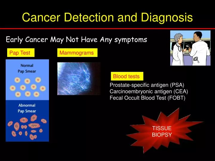
Cancer Detection and Diagnosis. Pap Test. Mammograms. Blood tests. Mammograms. Mammograms. Mammograms. Mammograms. Prostate-specific antigen (PSA) Carcinoembryonic antigen (CEA) Fecal Occult Blood Test (FOBT). Early Cancer May Not Have Any symptoms. TISSUE BIOPSY. Tumour grading.

E N D
Cancer Detection and Diagnosis Pap Test Mammograms Blood tests Mammograms Mammograms Mammograms Mammograms Prostate-specific antigen (PSA) Carcinoembryonic antigen (CEA) Fecal Occult Blood Test (FOBT) Early Cancer May Not Have Any symptoms TISSUE BIOPSY
Tumour grading Microscopic examination - - likely behavior - responsiveness to treatment. "grade" a low number grade (grade I or II) refers to cancers with fewer cell abnormalities than those with higher numbers (grade III, IV).
Tumour Staging • How large is the tumour, and how far has it invaded into surrounding tissues? • 2. Have cancer cells spread to regional lymph nodes? • 3. Has the cancer spread (metastasized) to other regions of the body?
Tumour Suppressor Genes P53 Rb
Retinoblastoma Rare childhood cancer of the eye that develops in children, typically under five years old. Incidence • 2 % of childhood malignancies Influencing factors 30-40% hereditary 60-70% sporadic Treatment Surgery, radiation, chemotherapy
Retinoblastoma protein (pRb) • Normally inhibits cell proliferation • localised in the nucleus • tumour suppressor protein of ~110kD • pRb has > 10 phosphorylation sites (affects protein-protein interaction) • Rb gene is 300kb long & mutations in this gene leads to loss of function. • Most mutations involve gross chromosomal changes in the 3kb coding region and 1/3 are point mutations. • Loss of heterozygosity at chromosome 13q14.2.
p53 – guardian of the genome • Initially identified as a tumour specific nuclear antigen with a Mol wt of 53kDa • Comparison with normal cells showed the presence of mutations in cancer cells • When wild-type gene transfected into tumours, it stopped their growth • i.e. a tumour suppressor gene
p53 – guardian of the genome • 50% of all cancers show mutations in p53 • 90% mutations in Squamous Cell Carcinoma (SCC) • 80% point mutations and 20% truncations • Mutations cause loss of function • Leads to continued cell division despite having DNA damage • Leads to increased mutation rate mutant Wild type
p53 – guardian of the genome Cellular stress Cell proliferation Stimulates DNA repair Apoptosis
P53 – domain structure Transactivation domain Tetramer formation Auto inhibition DNA binding 300 100 200 393 1
P53 – Transcriptional activation Transactivation domain Tetramer formation Auto inhibition DNA binding stimulates transcription indirectly by binding to other nuclear proteins e.g.Mdm2, GADD45, Cyclin G, BAX, IGF –BP3
P53 – transcriptional activation DNA damage Cell proliferation Mdm2 Mdm2 P ARF transcription degradation
P53 – DNA binding Transactivation domain Tetramer formation Auto inhibition DNA binding Sequence specific DNA binding: Certain genes have a p53 response element that specifically binds to the p53 tetramer e.g. BAX, p21
P53 – mutational hotspots The C-terminal regulatory domain has 2 functions Negative regulation: Phosphorylation destabilises the folding of the DNA binding domain Positive regulation:Acetylation of the C-terminus of DNA-bound p53 stabilises p300 binding, which is required for p53 driven transcription
P53 – tetramer formation Transactivation domain Tetramer formation Auto inhibition DNA binding 300 100 200 393 1
P53 – Autoinhibitory domain Transactivation domain Tetramer formation Auto inhibition DNA binding Causes transcriptional repression e.g. JUN, FOS, PCNA, MYC, BCL2 genes