Download
1 / 35
380 likes | 581 Views
Vision. “El ojo que ves no es ojo porque tu lo veas, es ojo porque te ve ” Antonio Machado “The eye you see is not an eye due to you seeing it, It’s an eye because it sees you”. Vision: Outline. Light Eye Visual Path Visual Cortex. UV rays. Wave frequency. Wave

E N D
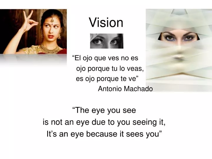
Vision “El ojo que ves no es ojo porque tu lo veas, es ojo porque te ve” Antonio Machado “The eye you see is not an eye due to you seeing it, It’s an eye because it sees you”
Vision: Outline • Light • Eye • Visual Path • Visual Cortex
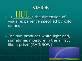
UV rays Wave frequency Wave amplitude Purity of the wave Perceptual Dimensions of Light
The Eye Cornea Pupil/Iris Lens Retina Cones Rods Fovea Optic disc (blindspot) 4
Similarity btw eye & camera known since 1600’s Eye anatomy: Functions transparent medium air (cornea, aqueous humor, pupil, lens, vitreous humor) lens lens iris diaphragm retina film a focal point a focal point
Eye Anatomy: Abnormalities Near-sightedness (Myopia ): image falls too short of retina (eyeball too long) newborns Far-sightedness: focal point of light falls beyond retina (Eyeball is too short) Lasik Changes the shape of the cornea (Laser-Assisted In Situ Keratomileusis)
Near-Sightedness nearby things are on focus
Cataracts • Reduced illumination, acuity, and color saturation • Deposits in the lens • Common in older adults
Eye Anatomy: Retina Red eye in photos due to dilated pupils Retina of diabetic patient • fovea: center of the retina, high concentration of cones • optic disk (blindspot) & direct view of arteries (clinical importance) • photorreceptors: cones (color vision) and rods
Concentration of Cones & Rods in Retina Visual Acuity Eye Anatomy: Retina
One Cones --> one ganglion cell high acuity (fovea) Many Rods --> ganglion cells. High sensitivity (periphery) (e.g, night vision)
Eye Anatomy: Optic disc (blindspot) Lateral visual field Medial Retina The eye is a device 'designed' to see, but the ‘blindspot’ reveals it is not perfect
Receptive field (RF) • is that portion of the visual field (outside world) in which the presentation of visual stimuli will produce an alteration in the firing rate of a particular neuron
Peak sensitivities of the three cones Tri-chromatic theory • Blue, red, & green “color” receptors COLOR VISION
No ‘green’ cones Test for Deuteranopia: Name number: (‘5’ or ‘2’) If you see a 2: Red/Green Color blindness (male) Most people who are color blind can see colors
Vision: Outline • Light • Eye • Visual Path & its deficits • Visual cortex
Hemianopia – objects are bisected with ½ obscured experiencing the obscured part as “blank” or “void” Scotoma: A small blindspot in the visual field caused by a small lesion, usually in the occipital lobe
Vision: Outline • Light • Eye • Visual Path & its deficits • Visual cortex • V1: Orientation sensitive • Ventral Pathway • Dorsal Pathway
Visual Cortex V1: primary visual cortex
V1 cells respond to lines of particular orientations of particular widths. Primary visual cortex (V1)
Vision: Outline • Light • Eye • Visual Path & its deficits • Visual cortex • Orientation sensitive • Ventral Pathway • Area MT (motion), Object Recognition, Area V4 (color) • synesthesia • Dorsal Pathway • Spatial Attention • Hemispatial Neglect
Cortical Connections of Visual areas • Complex & with multiple connections • Over-simplified version: dorsal & ventral paths
Ventral & Dorsal Paths ¼ of the brain is involved in visual processing, more than for all other senses
& how Ventral & Dorsal Paths
Ventral Path: Object recognition Lesion of ventral pathway Agnosia fMRI: Object recognition
Ventral Pathway (V4): Color perception Cerebral Achromatopsia: bilateral damage to V4 Color is more important of ‘what’ than for ‘where’ Synesthesia
Ventral Path: Objects vs. Faces Are faces very difficult objects or special ones (i.e., specific process)
Cars-Objects Birds-Objects Car Experts Fusiform Gyrus Bird Experts Gauthier et al., 2000 Fusiform Gyrus Neuroimaging of face, bird and car experts Fusiform Gyrus “Face Experts”
Fusiform Gyrus Fusiform Gyrus Children with autism as face “novices” Faces Control Group Autism Group Hypoactivation of fusiform face area Schultz, et al. 2000
In sum, different parts of the visual cortex are specialized in the processing of specific features • For example, • movement, • color. • Objects • Faces • Location Binding problem: If the brain processes features separately, how does it bind those features into a single conscious representation: Answer: Attention