Download
1 / 43
430 likes | 443 Views
Blood/Lymphatic Disorder Chapter 34. White Christensen Adam. L. Lehmkuhl, 2009. Blood-viscous red fluid containing RBCs, WBCs, and platelets in fluid called plasma Slightly alkaline with pH 7.35 to 7.45 NaCl concentration 0.9% 5-6 liters. Functions of blood

E N D
Blood/LymphaticDisorderChapter 34 White Christensen Adam L. Lehmkuhl, 2009
Blood-viscous red fluid containing RBCs, WBCs, and platelets in fluid called plasma Slightly alkaline with pH 7.35 to 7.45 NaCl concentration 0.9% 5-6 liters Functions of blood Transports O2 and nutrition to the cells and waste products away from the cells via cardiac and respiratory systems. Regulates acid-base balance, aids in temp regulation, and controls water content Protects against infection Blood/LymphaticDisorder
Erythrocytes (RBCs) Hgb 14-18 g/dl in males, 12-16 g/dl in females Lifespan 120 days Produced in red bone marrow Production stimulated by Erythropoietin Hct 42-52% in males, 37-47% in females Leukocytes (WBCs) Unlike RBCs, have a nucleus Fight infection and assist in immunity. 5,000-10,000 Elevated WBC’s indicate infection, inflammation, tissue necrosis or leukemia. Different kinds of WBCs are counted and reported as percentages of the total is called a differential 2 categories- granulocytes and agranulocytes Wright’s stain differentiates category of WBC Blood/Lymphatic Disorders
Granulocytes Neutrophils- essential for phagocytosis (process of engulfing) Immature polymorphonuclear leukocytes called bands are released into the blood stream when neutrophils are depleted and severe infection occurs Eosinophils- play a role in allergic reactions and against certain parasitic worms Basophils- nonspecific immune response, release histamine Blood/LymphaticDisorders
Agranulocytes Monocytes- function similarly to neutrophils through process of phagocytosis Lymphocytes (B & T)- Blood/LymphaticDisorders
Thrombocytes (platelets) Nonnucleated Life span of about 10 days. Produced in red bone marrow Function in process of hemostasis (prevention of blood loss) and in clotting formation Hemostasis Vessel spasm Platelet plug formation Clot formation Blood Types A, B, AB, and O Type O is the universal donor(no antigens that bodies can attack) Type AB is the universal recipient Rh factor- 85% of humans carry this factor Rh incompatibility seen most often in pregnancy causing hemolysis of the RBCs causing rupture and loss of cell contents Blood/LymphaticDisorders
The complete blood count (CBC) is a screening test, used to diagnose and manage numerous diseases. It can reflect problems with fluid volume (such as dehydration) or loss of blood. It can show abnormalities in the production, life span, and destruction of blood cells. It can reflect acute or chronic infection, allergies, and problems with clotting. Complete Blood Count -
A CBC requires a small blood specimen. Blood is drawn from a vein, usually from the inside of the elbow or the back of the hand. Preparation: The skin should be cleaned with alcohol or iodine before the test. The patient should be seated comfortably or reclining. Complete Blood Count
Low numbers of red blood cells may indicate anemia, Blood loss Iron deficiency Deficiences of vitamin B12 or folic acid Bone marrow failure (for example, from radiation, toxin, fibrosis, tumor) Erythropoietin deficiency (secondary to kidney disease) Hemolysis (RBC destruction) Leukemia Multiple myeloma Over hydration Complete Blood Count Normal values vary with altitude and gender.
High numbers of red blood cells may indicate: Cigarette smoking Low oxygen tension in the blood Congenital heart disease Cor pulmonale Pulmonary fibrosis Polycythemia vera Dehydration (such as from severe diarrhea) Emphysema Complete Blood Count
Low numbers of white blood cells (leukopenia) may indicate: Bone marrow failure (for example, due to granuloma (granular tumor), tumor, or fibrosis) Presence of cytotoxic substance Collagen-vascular diseases (such as lupus erythematosus) Disease of the liver or spleen Radiation exposure High numbers of white blood cells (leukocytosis) may indicate: Infectious diseases Inflammatory disease (such as rheumatoid arthritis or allergy) Leukemia Severe emotional or physical stress Tissue damage (for example, burns) WBC’s
Whole blood (^ vol) Packed RBC’s (anemia) Platelets (control bleeding) Plasma (clotting d/o’s) Cryoprecipate (fibrinogen deficiencies) Administer within 30 minutes. Complete within 4 hours. Baseline vs 18-19 guage catherter 2 nurse check Anyreaction immediately stop infusion..call MD N/S to prevent clotting Autologous or homologous Blood Transfusions
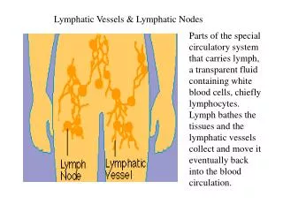
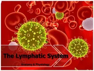
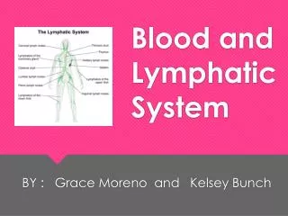
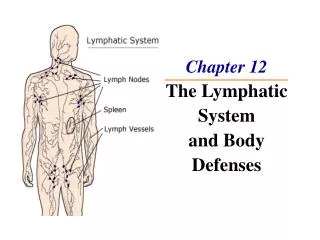
Lymphatic system- consists of lymph vessels, lymph fluid, and lymph tissue 2 main functions Maintenance of fluid balance (excess fluid from interstitial space to circulatory system) Production of lymphocytes to protect body from infection. Lymph nodes- filter impurities from the lymph and produce lymphocytes and macrophages Superficial lymph nodes in neck, axilla and groin can be palpated, especially when infected and swollen. CA cells can collect, reproduce and travel through lymph. Lymph nodes are biopsied to detect spread of CA. Blood/LymphaticDisorders
Tonsils- lymphoid tissue in the oropharynx Spleen- organ located in the LUQ of the abdomen, contains lymphatic nodules, stores 350 ml of blood. If needed approx. 200mL can be pumped out within a minute as needed. Removes old RBC’s, platelets and microorganisms from blood. During infection spleen enlarges to produce and release monocytes and lympthocytes. Thymus- located in upper thorax, develops T lymphocytes (Tcells). Large in infancy and childhood Decreases in size with age T cells are actively involved in immunity. Blood/LymphaticDisorders
Diagnostic tests CBC Red cell indices- MCV (size), MCH (weight), MCHC (concentration) Peripheral smear- most informative test Schilling- measures the absorption of radioactive vitamin B12 in diagnosing pernicious anemia Gastric analysis- useful in determining pernicious anemia Lymphangiography- detects metastatic involvement of lymph nodes Bone marrow aspiration or biopsy- most commonly performed in persons with marked anemia, neutropenia, acute leukemia, and thrombocytopenia Blood/LymphaticDisorders
Anemia- disorder characterized by low RBC, Hgb, Hct, and RBC destruction Hypovolemic anemia (blood loss) Secondary anemia due to blood loss Control bleeding, tx shock, replace fluid volume, O2 Pernicious anemia Absence of intrinsic factor produced by gastric mucosa which is needed for absorption of B12 Schilling test, serum B12 test, gastric analysis Tx is B12 injections (for life), folic acid Pt is highly susceptible to gastric carcinoma and should be monitored closely for symptoms. Blood/LymphaticDisorders
Aplastic anemia Failure of the bone marrow to produce RBCs Patient may have pancytopenia (low WBC, RBC,platelets) Most cases the cause is unknown..maybe genetic, or secondary to viral invasion, medications, chemicals, radiation, or chemotherapy Bone marrow transplant, if pts bone marrow fails to respond to tx. Prevent infection, bleeding, and fatigue For transplant the best candidate is a young pt who has not had previous infusions. Sibling donor under 30 yrs is higher success Immunosupressants are given to prevent graft rejection. Blood/LymphaticDisorders
Iron deficiency anemia Most common type of anemia. RBCs contain decreased Hbg Most common cause is chronic intestinal or uterine bleeding Low RBC, Hgb, Hct, and serum iron levels May have pica, stomatitis, glossitis, and brittle hair if chronic. Population with higher incidence are: Low birth wt infants or premature Infants Adolescent girls Alcoholics Menstruating women Pregnancy Tx is with iron salts such as Ferrous Sulfate Ascorbic acid enhances iron absorption Z track if parenteral Use straw with liquid Makes stools green or black Diet in Fe rich foods Blood/LymphaticDisorders
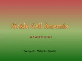
Sickle cell anemia is caused by an abnormal type of hemoglobin called hemoglobin S. Hemoglobin is a protein inside red blood cells that carries oxygen. Hemoglobin S, however, distorts the red blood cells' shape. The fragile, sickle-shaped cells deliver less oxygen to the body's tissues, and can break into pieces that disrupt blood flow. Sickle cell anemia is inherited as an autosomal recessive trait. This means it occurs in someone who has inherited the hemoglobin S gene from both parents. Sickle cell is the mostcommon genetic D/O in the US particularly in African-Americans,,Asian, India, Mediterranean and Caribbean areas. Sickle Cell Anemia
1 in 10 African Americans have the trait, 1 in 500 have the disease Dx with Hgb electrophoresis which shows HbS Negative means sickle cell trait not sickle cell disease. Individual is asymptomatic but may pass disease to offspring. If Hbss (genes) confirmed dz No specific treatment teach to avoid sickle cell crisis Sickle cell crisis manifestations: Fever Severe pain Loss of blood to various organs (obstructed vessels) Areas most affected are joints (become painful and swollen), bone, brain, lung, liver, kidneys and penis. Frequent PVS, assessment, heat to joints, avoid tight clothing and high altitudes. Tx symptoms Hydroyurea (pain),folic acid (^RBC production) PCA during crisis, ^HOB, O2 ^ tissue perfusion and offset dyspnea) Sickle Cell
Two types Primary and Secondary (compensory mech to make more rbcs secondary to hypoxia from smoking, living in high altitude, HF) Primary- Jewish men >50 y/o acquired mutation (abnormal DNA) occurs in bone marrow. Erythrocytosis in Polycythemia Vera Hypervolemic, hyperviscous state (blood is unable to circulate freely) Increased plasma, RBC (WBC’s and platelets also increased), Hbg, Hct, low oxygen level on ABG Tx is repeated Phlebotomy (350-500mL) to decrease RBC’s, blood viscosity and reduce blood volume. Myelosuppressive agents- Myleran, hydroxyurea, and radioactive phosphorous (to decrease RBC production) Low Na+ diet (decrease blood vol and avoid Fe rich foods Polycythemia
Malignant D/O with excess WBCs in marrow and lymph nodes Pt constantly fighting infections and has fever and chills. Enlarged lymph nodes, anemia, thrombocytopenia (bleeding, bruising), low or very high WBCs Hepatosplenomegaly, lymphadenopathy, bone pain (WBC;s crowding cells in bone marrow), oral lesions Bone marrow bx shows immature WBCs Bleeding common monitor platlets Tx is chemo + radiation (destroys CA cells and good cells), bone marrow transplant Prevent infection (high risk), and chronic pain Most common cause of death is viral or fungal PNA. Acute lymphocytic leukemia (ALL) <15 yo more rapid onset than Acute myeloid leukemia (AML). R/f infection and bleeding Teach: Hand washing to visitors, pt hygiene use antimicrobial soaps, electric razor, antiemetics, stool softener, monitor for hematuria or cloudy urine. Leukemia
Severe reduction in granulocytes (basophils, eosinophils, and neutrophils) Leukopenia Med reaction or toxicity, cancer, chemotherapy or radiation Differential below normal Tx is to alleviate cause and to prevent infection Meticulous handwashing, monitor: VS, WBC, lung sounds. Strict aseptic technique, visitors screened, reverse isolation may be implemented. Agranulocytosis
Coagulation Disorders • Disseminated Intravascular Coagulation • Hemophilia • Thrombocytopenia
Inherited blood clotting d/o from lack of certain clotting factors Hemophilia A most common Hereditary, x linked, affects mostly males, females carry genes Disturbance in clotting factors Dx Factors VIII and IX, prolonged PTT Tx transfusions, replacement of factor VIII and IX (cryoprecipitate) Willebrand’s Disease- mild deficiency of factor VIII 3 classifications Mild Moderate Severe Can bleed out from trauma or bleed spontaneously. Spontaneous ecchymosis from GI or GU tracts Hemophilia
Acquired Hemorrhagic syndrome of the clotting cascade and over stimulation of the clotting process. DIC a condition of alternating clotting and hemorrhaging. Coagulopathy resulting from the overstimulation of clotting and anticlotting processes in response to disease or injury Dx prolonged clotting coagulation profile, marked thrombocytopenia, deficits in factor V and VIII Tx cryoprecipitate, heparin, fibrinolytics, transfusion Primary dz stimulates the clotting mechanism , causes mini microthrombi to develop. The body’s fibronolytic process attempts top break clots thus causing hemorrhaging. Disseminated Intravascular Coagulation (DIC)
Platelets reduced below 100,000 Drug induced, infection or idiopathic Risk for bleeding Prevent trauma (0 falls) or infection Petechiae (small skin hemorrhages) and ecchymosis (bruising). Monitor for internal bleeding Tx corticosteroids and splenectomy (last resort..spleen is where platelet destruction occurs), platelet transfusions or apheresis (removal of unwanted componenets), IV gamma globulin or immunosuppressive drugs Thrombocytopenia
Lymph Disorders • Hodkins Lymphoma • Non-Hodkins Lymphoma
Malignant Lymphoma Cancer of lymphoid tissue Painless, enlarged lymph nodes, fever, wt loss, anemia, pruritus, and prone to infection Bone scan may reveal fxs from bone mets, CT scan, node bx Tx chemo and radiation Supportive care, interventions similar to Hodgkins Lymphoma Hodgkins Lymphoma Cancer of the lymphoid tissue Males, bimodal Biopsy of lymphoid tissue shows Reed Sternberg Cells. Tx dependent on stage Tx chemo and radiation Nursing care according to stage Comfort measures focus on skin integrity Soothing baths with antipruritic medications (e.g. puritis). Blood/LymphaticDisorders
Plasma Cell Disorder • Myeloma
Malignant neoplastic immunodeficiency disease of the bone marrow Plasma cell tumor Disruption of production of RBC, WBC, and platelets due to overproduction of plasma cells Bone destruction with release of calcium and phosphorous from bones Hyperuricemia with high Pro leads to renal failure Pain relief, prevent infection and bone injury, chemo, and hydration Encourage 3-4 Lof H2O to minimize complications of excessive Ca in the blood and urine. Exercise to preveny bone demineralization Monitor for hypercalciumia Multiple Myeloma