Download
1 / 72
730 likes | 913 Views
Chapter 27 Prokaryotes and the Origins of Metabolic Diversity. I. The world of prokaryotes A. They’re everywhere! 1. Collective prokaryote biomass outweighs all eukaryotes combined by at least tenfold.

E N D
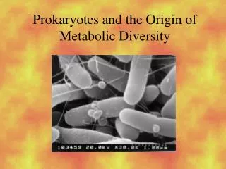
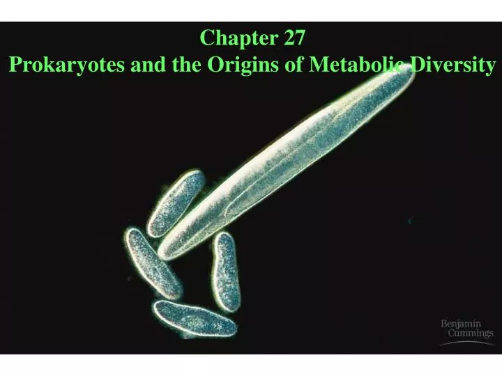
Chapter 27 Prokaryotes and the Origins of Metabolic Diversity
I. The world of prokaryotes A. They’re everywhere! 1. Collective prokaryote biomass outweighs all eukaryotes combined by at least tenfold. 2. They exist almost everywhere, including places where eukaryotes cannot. Figure 27.1 (p. 527) – “Heat-loving” prokaryotes.
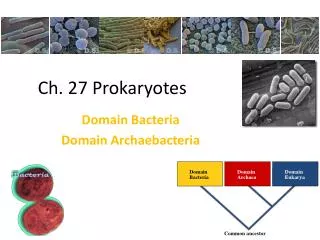
3. Most prokaryotes are beneficial; we couldn’t live without them. (e.g. Nitrogen-fixing bacteria) 4. Some cause illness à bubonic plague, diphtheria, salmonella 5. Approximately 5000 species have been identified. Estimates of prokaryote diversity range from 400,000 to 4,000,000 species. B. Bacteria and archaea are the two main branches of prokaryote evolution 1. Archaea are thought to be more closely related to eukaryotes than to bacteria. Figure 27.2, p. 527, Ed. 6) – The three domains of life. Figure 27.12, . 540, Ed. 7
II. Structure, function, and reproduction of prokaryotes A. Most prokaryotes are unicellular. 1. Some species form aggregates of two or more individuals. B. Three (3) common shapes: cocci (round); bacilli (rod); helical (spiral) Figure 27.3 (p. 528, edition 6) – The most common shapes of prokaryotes. Figure 27.2 (p. 535, edition 7)
C. Prokaryotes are typically 1-5 μm in diameter, but some can be seen by the naked eye. - Eukaryotic cells are typically 10-100 μm in diameter. Figure 27.4 (p. 528) – The largest known prokaryote.
D. Almost all prokaryotes have cell walls external to the plasma membrane. 1. Cell walls maintain cell shape. 2. Cell walls are composed of peptidoglycan.
3. There are two types of cell walls. Bacteria are grouped according to cell wall type. a. Gram-positive bacteria have simple, thick cell walls. Their cell walls are composed of a relatively large amount of peptidoglycan. b. Gram-negative bacteria have less peptidoglycan and are more complex. They have a peptidoglycan layer surrounded by the plasma membrane and an outer membrane. - Gram-negative bacteria are typically more resistant to host immune defenses and antibiotics. Note that the two types of bacteria can be stained to determine which is gram-negative (pink) and gram-positive (purple) using a Gram Stain. Figure 27.5 (p. 529, Ed. 6) – Gram-positive and gram-negative bacteria. (Fig. 27.3, Ed. 7)
4. Most prokaryotes secrete sticky substances that form a protective layer that enables them to adhere to substrates. a. The sticky protective layer secreted by prokaryotes is called the capsule. 5. Some prokaryotes adhere to substrates using pili and fimbriae. a. Some pili are specialized for DNA transfer. This process is called conjugation; note for later in class. Figure 27.6 (p. 530, Ed. 6) – Pili.
E. Many prokaryotes are motile - Some exceed speeds 100 times their body length per second. 1. Modes of movement – Note the three types: a. Flagellum- basal apparatus rotates the flagellum and propels the cell Figure 27.7 (p. 530, Ed. 6) – Form and function of prokaryotic flagella. Figure 27.6, p. 536, Ed. 7 b. Corkscrew movement of spirochetes (helical) c. Some prokaryotes glide over jets of slimy secretions. 2. Many prokaryotes move toward or away from a stimulus àtaxis. Chemotaxis is the movement toward or away from a chemical.
F. Cellular and genomic organization of prokaryotes is different from that of eukaryotes 1. Prokaryotes have no nucleus. 2. The nucleoidregion in a prokaryotic cell consists of a concentrated mass of DNA. This mass of DNA is usually one thousand times less than what is found in a eukaryote. 3. A prokaryote may have a plasmid in addition to its major chromosome. A plasmid is a small ring of DNA that carries accessory genes. Usually these genes are for antibiotic resistance!
G. Prokaryotes grow and adapt rapidly- The doubling time for E. coli is 20 minutes. Start with one E. coli cell. After 48 hours of doubling every 20 minutes, the mass of E. coli would be 10,000 times the mass of the earth. Bacteria do not have gene transfer by sexual reproduction, but do transfer genes. Why? This is an aid in adapting (evolving). 1. Three (3) ways for genes to be transferred between cells: a. Transformation – cell takes up genes from the surrounding environment. b. Conjugation – direct transfer of genes from one prokaryote to another. Use the sex pilus to conjugate. c. Transduction – viruses transfer genes between prokaryotes.
2. Endospores are resistant cells formed by some bacteria as a way to withstand harsh conditions. The cell replicates its chromosome and wraps it in a durable wall that can protect the chromosome from adverse conditions, e.g. boiling water, desiccation. When the environment is good again, the cell will revive to a new vegetative (growing) state. Figure 27.10 (p. 532) – An anthrax endospore.
III. Nutritional and metabolic diversity A. All prokaryotes (and eukaryotes too) are grouped into four (4) categories according to how they obtain energy and carbon Table 27.1 (p. 533, Ed. 6; p. 339, Ed. 7) – Major Nutritional Modes 1. Photoautotrophs - Photosynthetic à use light as the energy source - CO2 is the carbon source Example: Cyanobacteria; plants (eukaryotic). Figure is of Anabaena – Genus of photosynthetic cyanobacterium that fixes N2 in specialized cells called heterocysts A major source of organic nitrogen in the world.
2. Chemoautotrophs - Energy from oxidation of inorganic substances (e.g. NH4, and S) - CO2 is the carbon source Example: Sulfolobus, Beggiatoa (shown on slide)
3. Photoheterotrophs - Light as energy source - Organic compounds are source of carbon 4. Chemoheterotrophs - Organic compounds are energy source and source of carbon (this includes humans) Examples: Many prokaryotes; animals (eukaryotic); fungi (eukaryotic)
B. Metabolic relationships to oxygen 1. Obligate aerobes - Use O2 for respiration; cannot grow without it. (Humans are obligate aerobes) 2. Facultative aerobes - Use O2 when available; ferment when O2 isn’t available. 3. Obligate anaerobes - Poisoned by O2; use fermentation or live by anaerobic respiration. In anaerobicrespiration, inorganic molecules like SO4, NO3, and Fe3+ are used instead of oxygen.
C. Photosynthesis evolved early in prokaryotic life 1. Cyanobacteria started to produce O2 about 2.7 billion years ago
IV. A survey of prokaryotic diversity Figure 27.13 (p. 536, Ed. 6) – Some major groups of prokaryotes. Figure 27.12, p. 540, Ed. 7.
A. Great diversity of Archaea in extreme environments and oceans 1. Two taxa of archae: a. Euryarchaeota – most archae b. Crenarcheota – most thermophilic species 2. Examples of extremophiles a. Methanogens produce methane - Energy is from hydrogen gas - Strictly anaerobic - Inhabit swamps and animal intestines b. Extreme halophiles - Live in salty environments (Great Salt Lake) Figure 27.14 (p. 537) – Extreme halophiles.
c. Extreme thermophiles - 60- 80 °C optimum temperatures (hot springs) - 105 °C for deep-sea hydrothermal vents
B. Most prokaryotes are in the Domain Bacteria. Table 27.3 (p. 537) – A Comparison of the Three Domains of Life - There are five clades to remember: 1. Proteobacteria - Gram-negative bacteria - Broad, diverse group that has five subgroups REMEMBER THE FOLLOWING BACTERIA WITHIN THIS CLADE AND WHAT THEY DO!!!