Download
1 / 25
250 likes | 362 Views
Skeletal Cartilages. Contain no blood vessels or nerves Dense connective tissue girdle of perichondrium contains blood vessels for nutrient delivery to cartilage. Epiglottis. Larynx. Thyroid cartilage. Cartilage in external ear. Cartilages in nose. Trachea. Cricoid cartilage. Lung.

E N D
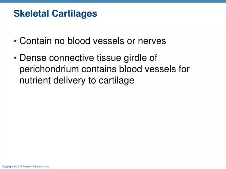
Skeletal Cartilages • Contain no blood vessels or nerves • Dense connective tissue girdle of perichondrium contains blood vessels for nutrient delivery to cartilage
Epiglottis Larynx Thyroid cartilage Cartilage in external ear Cartilages in nose Trachea Cricoid cartilage Lung Articular Cartilage of a joint Cartilage in Intervertebraldisc Costal cartilage Respiratory tube cartilages in neck and thorax Bones of skeleton Pubic symphysis Axial skeleton Meniscus (padlike cartilage in knee joint) Appendicular skeleton Cartilages Articular cartilage of a joint Hyaline cartilages Elastic cartilages Fibrocartilages Figure 6.1
Cartilage in external ear Cartilages in nose Articular Cartilage of a joint Cartilage in Intervertebraldisc Costal cartilage Pubic symphysis Meniscus (padlike cartilage in knee joint) Articular cartilage of a joint Figure 6.1
Functions of Bones • Support • Protection • Movement • Storage • Minerals (calcium and phosphorus) • Blood cell formation in marrow cavities
Articular cartilage Compact bone Proximal epiphysis Spongy bone Epiphyseal line Periosteum Compact bone Medullary cavity (lined by endosteum) (b) Diaphysis Distal epiphysis (a) Figure 6.3a-b
Membranes of Bone • Periosteum • Outer fibrous layer • Inner osteogenic layer • Osteoblasts (bone-forming cells) • Osteoclasts (bone-destroying cells) • Nerve fibers, nutrient blood vessels, and lymphatic vessels enter via nutrient foramina • Secured to underlying bone by Sharpey’s fibers
Endosteum Yellow bone marrow Compact bone Periosteum Perforating (Sharpey’s) fibers Nutrient arteries (c) Figure 6.3c
Spongy bone (diploë) Compact bone Trabeculae Figure 6.5
Artery with capillaries Structures in the central canal Vein Nerve fiber Lamellae Collagen fibers run in different directions Twisting force Figure 6.6
Spongy bone Compact bone Central (Haversian) canal Perforating (Volkmann’s) canal Endosteum lining bony canals and covering trabeculae Osteon (Haversian system) Circumferential lamellae (a) Perforating (Sharpey’s) fibers Periosteal blood vessel Lamellae Periosteum Nerve Vein Lamellae Artery Central canal Lacuna (with osteocyte) Canaliculi Osteocyte in a lacuna Lacunae Interstitial lamellae (b) (c) Figure 6.7a-c
Nerve Vein Lamellae Artery Central canal Canaliculus Lacunae Osteocyte in a lacuna (b) Figure 6.3b
Chemical Composition of Bone • Organic bone matrix secreted by osteoblasts • Collagen fibers 33% • Provide tensile strength and flexibility • Inorganic hydroxyapatites (mineral salts) • 65% of bone by mass • Mainly calcium phosphate crystals • Responsible for hardness
Bone Development • Osteogenesis (ossification)—bone tissue formation • Stages • Bone formation—begins in the 2nd month of development • Postnatal bone growth—until early adulthood • Bone remodeling and repair—lifelong
Resting zone Proliferation zone Cartilage cells undergo mitosis. 1 Hypertrophic zone Older cartilage cells enlarge. 2 Calcification zone Matrix becomes calcified; cartilage cells die; matrix begins deteriorating. 3 Calcified cartilage spicule Osteoblast depositing bone matrix Ossification zone New bone formation is occurring. 4 Osseous tissue (bone) covering cartilage spicules Figure 6.10
Hormonal Regulation of Bone Growth • Growth hormone stimulates epiphyseal plate activity • Thyroid hormone modulates activity of growth hormone • Testosterone and estrogens (at puberty) • Promote adolescent growth spurts • End growth by inducing epiphyseal plate closure
Bone Deposit • Occurs where bone is injured or added strength is needed • Requires a diet rich in protein; vitamins C, D, and A; calcium; phosphorus; magnesium; and manganese
Control of Remodeling • What controls continual remodeling of bone? • Hormonal mechanisms that maintain calcium homeostasis in the blood • Mechanical and gravitational forces
Hormonal Control of Blood Ca2+ • Calcium is necessary for • Transmission of nerve impulses • Muscle contraction • Blood coagulation • Cell division
Calcium homeostasis of blood: 9–11 mg/100 ml BALANCE BALANCE Stimulus Falling blood Ca2+ levels Thyroid gland Osteoclasts degrade bone matrix and release Ca2+ into blood. Parathyroid glands Parathyroid glands release parathyroid hormone (PTH). PTH Figure 6.12
Bone Repair 1 2 3 4 Blood from ruptured blood vessels forms a clot surrounding the ends of the broken bone Healing begins when a callus of cartilage replaces the clot Bone gradually replaces the cartilage in the callus When mature bone completely replaces the callus and the original shape of the bone has been mostly restored, the fracture is healed large blood clot compact bone spongy bone