Download
1 / 35
360 likes | 646 Views
Physics of the Eye and Intralase Surgery. Sight and lenses. The eye is a series of lenses that refract and focus light. These lenses follow the same laws of physics that artificial lenses would. Lenses and focal points.

E N D
Sight and lenses • The eye is a series of lenses that refract and focus light. • These lenses follow the same laws of physics that artificial lenses would.
Lenses and focal points • The cornea and lens of the eye act as two convex lenses. In order to understand illness and disorders of the eye, we must understand how lenses and focal points work.
Lenses and focal points To find the focal point, distance of an image, or distance to the object you can use the thin lens equation. Light bounces off an object from a distance s1. This light passes through the lens to be focused on the other side. This image is real, inverted, and smaller, and is located at a distance of s2 from the lens. The thin lens equation
Lenses and focal points Focal Point A convex lens
Lenses and focal points • Changing the distance to an object, or changing the focal length of a lens, will result in a different focal point. • Changes of these kind in the eye are the causes of visual acuity loss.
Sight, it’s a light thing. • Light is reflected off of objects and enters into the eye. • Colors are the wavelengths that are not adsorbed by the substance.
Anatomy and Function of the Eye • As light enters the eye, it first passes through the cornea. Most of the refraction of light occurs here. The refractive index of the cornea is 1.38. Refractive index= Speed of light in vacuum Speed of light in substance
Anatomy and Function of the Eye • The eye can control the amount of light that enters by the opening and closing pupil aperture using the iris.
<- Dilated Constricted ->
Anatomy and Function of the Eye • After passing through the cornea, light enters and refracts again through the lens of the eye. The lens has a refractive index of 1.44.
Anatomy and function of the eye This also changes the bulge of the cornea somewhat. This also aids to adapt vision into different situations. The eye can change the shape of the lens using muscles called ciliary muscles that stretch and flatten the lens, or relax and allow it to return to normal shape. This is called accommodation. Sometimes this bulging of the cornea is an appreciable change that can readily be seen by the naked eye.
Anatomy and Function of the Eye • The light then is focused onto the fovea, a cone and rod dense area on the retina, that produces the sharpest visual image.
Vision Disorders Normal eye. Focus hits directly on fovea to produce sharp image.
Vision Disorders This is known as nearsightedness, or myopia. The image comes into focus before the retina. Things that are near to the eye can more easily be seen. This condition can result from the eye being too long, the cornea being too round, or the lens having too much focus power.
Vision Disorders This condition is known as farsightedness, or hyperopia. The image produced by the lens and cornea comes into a focal point behind where the retina is located. Things that are farther away can be more easily seen. This results from the eye being too short, the cornea being too flat, or the focus power of the lens being too weak.
Vision Disorders This is a condition known as astigmatism. The shape of the lens and/or cornea is irregular leading to a loss of focal points or, more commonly, two focal points on the retina.
Vision Disorders This condition is known as presbyopia. The lens becomes rigid and fails to bend or accommodate to changing conditions.
Treatments • For centuries people have been using lenses to correct or compensate for vision loss. Traditional remedies are as follows:
Treatments A diverging lens will correct for nearsightedness. A converging lens corrects for farsightedness. Bifocal lenses will compensate for presbyopia. A custom ground lens made specifically for each eye will correct astigmatism.
Treatments • More recently, doctors have been using surgery to correct vision problems. These surgeries employ the same physics as corrective lenses would outside the eye, only the changes are done to the cornea and lens of the eye itself.
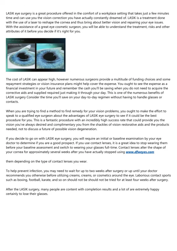
Treatments • Radial Keratotomy was one of the original vision correction surgeries. • Small cuts were made in the cornea radiating from the center. This was • used to treat mild cases of myopia. • Automated lamellar keratoplasty used a • microkeratome to make a small flap in the cornea. This was then folded out of the way, and the microkeratome was then used to sculpt the cornea. ALK can only correct myopia. A microkeratome
Treatments Photorefractive keratotomy is the precursor to LASIK surgery. This type of surgery was introduced in 1987. It uses a Eximer laser to reshape the outside of the cornea and thus correct vision. PRK can correct myopia, astigmatism, and some hyperopia.
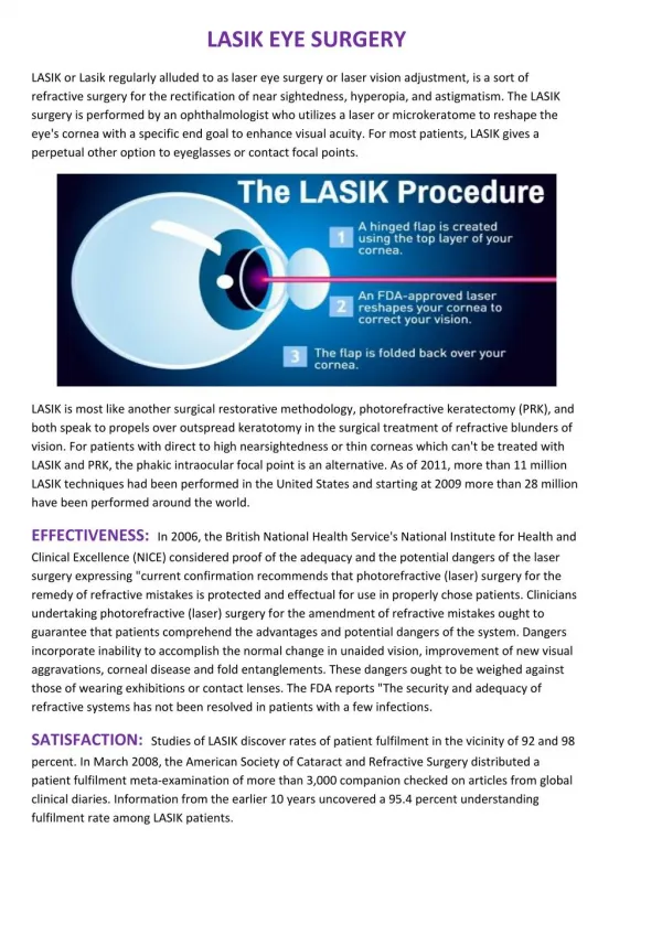
Treatments • In 1989 the FDA approved LASIK (Laser-Assisted in Situ Keratomileusis) • for treatment of: • Nearsightedness • Farsightedness • Astigmatism In LASIK surgery a mechanical cut is made across the cornea to create a flap of tissue and exposing the stroma of the cornea. This flap is moved aside and a “cool ultraviolet” laser sculpts and re-shapes the cornea, correcting acuity problems. This laser allowed for greater precision and thus greater flexibility for treatment of a broader range of symptoms.
Intralase Surgery In 2001, the FDA approved a laser to be used in a surgery called Intralase. Before surgery, exhaustive measurements are taken of each eye that will be operated on. Measurements of refractivity of the eye tell the prescription that each cornea and lens is contributing. The topography of the cornea and the measurements of the pupil are also taken.
Intralase Surgery The eye is anesthetized with drops and a suction ring is placed on the cornea to immobilize the eye. A guide cone is then placed on the ring to direct the eye appropriately. The laser will track the eye in case of accidental eye movement during the operation.
Intralase Surgery A femosecond laser then makes a very precise (+/- 10 microns) flap across the surface of the cornea, instead of a microkeratome. (A human hair is 50 microns in diameter). This reduces rare complications that LASIK presented, and also opened corrective eye surgery to previously ineligible patients. This laser produces carbon dioxide and water bubbles in the cornea. These bubbles are what actually produce the “incision” to create the flap. This can also be done vertically to produce a deeper incision.
Intralase Surgery • After waiting for the cavitation bubbles to disappear, the flap is pulled aside and the cornea is then sculpted to the needed measurements.
Intralase Surgery • The flap is then folded back, and the patient is ready to return home. • A few follow up appointments are done to ensure the eyes are moist and that the flaps are closed properly.
Intralase Surgery • Intralase and LASIK surgeries can repair hyperopia, myopia, and astigmatism. • By sculpting one eye to be “farsighted” and one to be “nearsighted”, you can eliminate the need for bifocals as well.
Intralase Surgery • Intralase surgery has the same benefits of LASIK, but with fewer risks by using a laser to make the flap instead of a mechanical cut. • Greater precision also allows patients that were at too high of a risk for complications before to now be eligible for surgery.
Bibliography Pictures: • http://sln.fi.edu/300/images/bifocals.jpg • plusinfo.jeonju.ac.kr/photos/Art • http://bnet.org/rbsd/ruhs/academics/science/physics/project2/thierren/convex.jpg • http://en.wikipedia.org/wiki/Lens_(optics) • http://www.shokabo.co.jp/sp_e/optical/labo/lens/lens_c2.jpg • http://www.eyeny.com/images/normal-vision.jpg • http://www.mcoaicare.com/images/eye.jpg • http://health.howstuffworks.com/lasik.htm • http://nate.calc.org/webalbum/fun • www.peterglass.com/funny%20glasses%20LG.htm • www.avclinic.com/rk.htm • www.institutdelamyopie.com/microkera.htm • www.focusmedical.ch/aserbehandlung_en.html • www.refraktives-zentrum.de/behandlung.php • 141.211.80.200/01regentsreport.html • www.uvcenters.com/refractive/lasik.html • www.intralaser.com/ • http://www.optometric.com/article.aspx?article=71162 • http://files.turbosquid.com/Preview/Content_on_5_8_2002_12_54_13/Lightbulb_Idea.jpg764383AE-CDAC-4977-AB1FEF08A38CBE19.jpgLarge.jpg
Bibliography Movies: • http://www.ebaumsworld.com/videos/popeyes.html • http://www.schustereyecenter.com/ts_main.sstg?page=visx References: • http://www.physicsclassroom.com/Class/refrn/U14L6a.html • http://www.durrievision.com/index.cfm/news/intralasedec • http://healthlink.mcw.edu/article/1031002532.html • http://www.optometric.com/article.aspx?article=71162 • http://health.howstuffworks.com/lasik.htm • http://www.fda.gov/cdrh/LASIK/what.htm • http://en.wikipedia.org/wiki/Lens_(optics)