Download
1 / 31
310 likes | 311 Views
This article explores the conduction of action potentials, including the role of currents, myelin sheath, and voltage-gated channels. It also discusses the cable theory of propagation and the different types of synapses involved in communication between neurons.

E N D
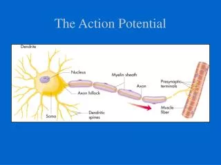
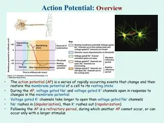
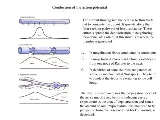
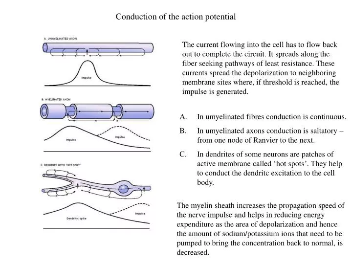
Conduction of the action potential The current flowing into the cell has to flow back out to complete the circuit. It spreads along the fiber seeking pathways of least resistance. These currents spread the depolarization to neighboring membrane sites where, if threshold is reached, the impulse is generated. • In umyelinated fibres conduction is continuous. • In umyelinated axons conduction is saltatory – from one node of Ranvier to the next. • In dendrites of some neurons are patches of active membrane called ‘hot spots’. They help to conduct the dendritc excitation to the cell body. The myelin sheath increases the propagation speed of the nerve impulse and helps in reducing energy expenditure as the area of depolarization and hence the amount of sodium/potassium ions that need to be pumped to bring the concentration back to normal, is decreased.
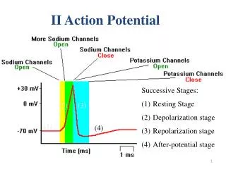
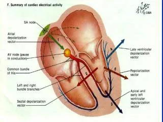
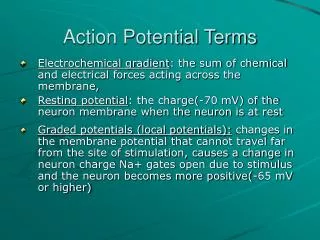
Unidirectional conduction of an action potential Unidirectional conduction of an action potential is due to transient inactivation of voltage-gated Na+ channels, which remain inactive for several milliseconds after opening. Reopening of Na+ channels “behind” the action potential is also prevented by the membrane hyperpolarization that results from opening of voltage-gated K+ channels
Channels distribution Sodium channels are dense at the node of Ranvier but sparse or absent in the internodal regions of the axon membrane. The K+ channels are located beneath the myelin sheath in internodal regions. There are about 700 000 sodium channels per node, i.e., 12,000 per um2 of nodal membrane. Internodal membrane can have no more than about 25 channels per um2.
Propagation of action potentials - cable theory To describe action potential propagation, it is necessary to derive the cable equation that illustrates how ions diffuse along the axons. In this respect the most useful geometrical structure is a cylinder. The parameters that are defined only for a cylinder are designated by small letters (ri, rm, cm) while parameters that are independent of any specific geometry will by designated by capital letters (Ri, Rm, Cm). ri rm, cm The definitions for the cylinder-dependent parameters are as follows: ri – axial resistance (W/cm) rm – membrane resistance (Wcm) cm – membrane capacitance (F/cm) ri corresponds to an infinitely thin disk of the cytoplasm with the same radius as the inside of the cylinder. rmand cm correspond to an infinitely thin ring of membrane, with the same radius as the cylinder. Extracellular resistance r0 = 0.
Propagation of action potentials - cable theory • The definitions for the membrane parameters independent of geometry are as follows: • Ri – specific intercelluar resistivity (Wcm) • Rm – specific membrane resistivity (Wcm2) • Cm – specific membrane capacitance (F/cm2) The membrane parameters are related to the cable specific parameters as follows: Total values of resistances and conductance (for a cable of length l) adding resistors in series adding resistors in parallel adding capacitors in parallel
Cable equations of action potential propagation The membrane current Im (uniform across the membranne) is given by: The decrease in Vm with distance is described by Ohm’s law: The decrease in ii with distance is equal to the current flowing across the membrane Action potentials propagate with a constant speed, so one can use the wave equation: We obtain: where, - conduction velocity (m/s)
Cable equations of action potential propagation From this wave equation, one can obtain: K = 10.47 m/s – estimated experimentally q= 18.8 m/s qexp= 21.2 m/s The Hodgkin and Huxley equations therefore give a very good fit to the experimental data
Two myths Neuron Electrical impulses Communication Electrical impulses komórka grzyba komórka jajowa strunowca komórka skóry żaby pień dyni komórka przysadki mógowej szczura komórka pierwotniaka komórka trzustki szczura
Synapsa Sir Charles Sherrington, 1897, Physiology textbook <gr. sýnapsis to clasp, connect or join>
Interneuronal relations Means of communication in the nervous system: Volume transmission - actions of neurotransmitters or neuropeptides at a distance, well beyond their release sites from cells or synapses Membrane juxtapositions – the membranes of two neruons are situated close together (~ 20 nm) Gap junctions – the membranes of two neruons are separated by a gap of 2-4 nm Chemical synapses – the most complicated and most characteristic. An example of membrane juxtapositions in a bundle of unmyelinated axons, which provide for interactions through ions (K+) or electric current (--). Types of junctions between nerve cells
Electrical and Chemical Synapses Distinguishing Properties of Electrical and Chemical Synapses Cytoplasmic continuity between pre- and postsynaptic cells Ultrastructural components Synaptic delay Type of synapse Distance between pre- and postsynaptic cell membranes Agent of transmission Direction of transmission Yes Gap-junction channels Virtually absent Electrical 3.5 nm Ion current Usually bidirectional No Presynaptic vesicles and active zones; postsynaptic receptors Significant: at least 0.3 ms, usually 1-5 ms or longer Chemical 20-40 nm Chemical transmitter Unidirectional
Electrical Synapses A. At electrical synapses two cells are structurally connected by gap-junction channels. A gap-junction channel is actually a pair of hemichannels, one in each apposite cell, that match up in the gap junction through homophilic interactions. The channel thus connects the cytoplasm of the two cells and provides a direct means of ion flow between the cells. This bridging of the cells is facilitated by a narrowing of the normal intercellular space (20 nm) to only 3.5 nm at the gap junction Electron micrograph: The array of channels shown here was isolated from the membrane of a rat liver. Each channel appears hexagonal in outline. Magnification × 307,800. B. Each hemichannel, or connexon, is made up of six identical protein subunits called connexins. C. The connexins are arranged in such a way that a pore is formed in the center of the structure. The pore is opened when the subunits rotate about 0.9 nm at the cytoplasmic base in a clockwise direction. Gap junctions in different tissues are sensitive to different modulatory factors that control their opening and closing. However, most gap-junction channels close in response to lowered cytoplasmic pH or elevated cytoplasmic Ca2+. • Main characteristics of electical transmission: • high speed • high fidelity • bidirectional • Functions: • extremely rapid transmission (e.g. tail-flip response) • synchronization of large group of neurons • communication in glial cells
Electrical synapses in Aplysia E. Kandel with Aplysia A train of stimuli applied to the tail produces a synchronized discharge in all three motor neurons. 1. When the motor neurons are at rest the stimulus triggers a train of identical action potentials in all three cells resulting in the release of ink. 2. When the cells are hyperpolarized the stimulus cannot trigger action potentials. Under these conditions the inking response is blocked.
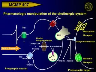
Chemical synapse • Action potential in nerve, depolarization of the terminal • Activation of voltage-gated Ca2+ channels • Fusion of vesicle to membrane • Release of neurotransmitter (exocytosis) • Diffusion of neurotransmitter across the synaptic cleft • Binding of neurotrasmitter to receptors and gating of ion channels. • Recycling of vesicles (endocytosis) • Inactivation of neurotransmitter
Patterns of synaptic connections Synapses (orange) in hippocampal cell
Acitve zones The active zone is the portion of the presynaptic membrane opposite the postsynaptic density across the synaptic cleft. The active zone is the site of synaptic vesicle docking and neurotransmitter release. Synapses (orange) in hippocampal cell Electron micrography of the synapse. Arrows point to the active zones – areas where synaptic vesicles in the nerve terminal cluster. Active zones are opposite a thickened, receptor-rich patch of postsynaptic membrane.
Neuromuscular junction Three neuromuscular junctions magnified 400X. Axon terminal [A] from a neuron is shown terminating into a large synaptic terminal [B] which communicates with a single skeletal muscle fiber. A muscle is innervated by a motor nerve fiber. The nerve fiber branches to form synaptic junctions with individual nerve fibers. Each junction (endplate) consists of a presynaptic nerve terminal from which acetylocholine is released, synaptic cleft, a postsynaptic area on the muscle containing receptors, and a surrounding envelope of glia. Electron microscope autoradiograph of the neuromuscular junction. T – axon terminal, M – muscle fiber. Scale 0.3 mm.
Neuromuscular junction – endplate potentials A. Intracellular recording of endplate potential (EPP) giving rise to an action potential (AP) in the muscle cell; experimental setup shown at left. B. High-gain recording showing summation of miniature endplate potentials (MEPPs). A single MEPP is due to a release of single quantum of ACh (~10000 molecules) at a single active zone (one quantum ~ one vesicle). Quanta released in synchrony by the impulse lead to summation of MEPPs and give rise to a large potential EPP. C. Very high gain recording showing noise induced by ionophopresis (using a small electric charge to deliver a chemical through the membrane) of ACh. D. Patch – clamp recording showing currents passing through single AChR channels.
Neuromuscular junction – endplate potentials A. Intracellular recordings from a muscle fiber at the endplate. B. The distribution of responses. The peaks in the histogram occur at amplitudes that are integral multiples of the amplitude of the unit potential (0.4 mV). This unit response is the same amplitude as the spontaneous miniature end-plate potentials (inset).
Quantal hypothesis (del Castillo, Katz, Martin) Neurotransmitter is released in quanta. Quantal release is a statistical, probabilistic, not a deterministic process. m = np m – number of quanta released n – number of possible quanta (roughly the number of active zones) p – average probability of release n ~ 1000 (number of active zones in a typical ending) m ~ 100-200 p ~ 0.1 – 0.2 The release of neurotransmitter in quanta applies to most of chemicalsynapses. The releaseprocess (probability) iscontrolled by the amount of depolarization of the nerve terminal membrane, whichinfluencescalciumlevel. The greater the Ca2+ influx into the terminal, the larger the number of quanta released.
The Ion Channel at the End-Plate Is Permeable to Both Sodium and Potassium The end-plate current is given by: I =g(V – VEPSP) I - the end-plate current, g – the conductance of the ACh-gated channels, V – the membrane potential,VEPSP – the chemical driving force, or battery. The ionic currents responsible for the end-plate potential can be determined by measuring the reversal potential of the end-plate current. The voltage of the muscle membrane is clamped at different potentials, and the synaptic current is measured when the nerve is stimulated. A. If Na+ flux alone were responsible for the end-plate current, the reversal potential would occur at +55 mV, the equilibrium potential for Na+ (VNa). The arrow next to each current record reflects the magnitude of the net Na+ flux at that membrane potential. B. The end-plate current actually reverses at 0 mV because the ion channel is permeable to both Na+ and K+, which are able to move into and out of the cell simultaneously. The net current is the sum of the Na+ and K+ fluxes through the end-plate channels. At the reversal potential (VEPSP) the inward Na+ flux is balanced by an outward K+ flux so that no net current flows.
Studying synapses Hippocampal slice in vitro chamber Cell culture Brain preparation in toto
The hippocampus is perhaps the most studied structure in the brain. It is critical to spatial learning and awareness, navigation, episodic/event memory and associational recollection. Hippocampus Sea horse (Hippocampus) The hippocampus is a part of the cerebral cortex, and in primates it is located in the medial temporal lobe, underneath the cortical surface The hippocampal Network: The hippocampus forms a principally uni-directional network, with input from the Entorhinal Cortex (EC, layers II-V) that forms connections with the Dentate Gyrus (DG) and CA3 pyramidal neurons via the Perforant Path (PP - split into lateral and medial). CA3 neurons also receive input from the DG via the Mossy Fibres (MF). They send axons to CA1 pyramidal cells via the Schaffer Collateral Pathway (SC), as well as to CA1 cells in the contralateral hippocampus via the Associational Commisural (AC) Pathway. CA1 neurons also receive inputs direct from the Perforant Path and send axons to the Subiculum (Sb). These neurons in turn send the main hippocampal output back to the EC, forming a loop. Entorhinal cortex - kora śródwęchowa, Subiculum - podkładka, Dentate gyrus - zakręt zębaty.
Excitatory synapses There are two main types of glutamate receptors: AMPA: α-amino-3-hydroxy-5-methyl-4-isoxazolepropionic acid NMDA: N-methyl-D-aspartate A. Intracellular recordings from a neuron responding to excitatory synaptic input at different holding potentials. The responses are shown before and after exposure to antagonists of the AMPA and NMDA receptors (APV is a NMDA receptor blocker). B. Diagrams showing AMPA and NMDA channels and the current flows through them.
Properties of NMDA receptors without Mg2+ with Mg2+ - NMDA receptor is voltage dependent due to voltage sensitivity of the Mg+ block.
Salwy epileptyczne in vitro RD Traub, JG Jefferys and MA Whittington. Enhanced NMDA conductance can account for epileptiform activity induced by low Mg2+ in the rat hippocampal slice, The Journal of Physiology, 1994, 478 (3) 379-393.
Inhibitory synapses There are two types of GABA (γ-aminobutyric acid)receptors: GABAA GABAB A. Intracellular recordings from a neuron responding to a sequence of excitatory and inhibitory synaptic inputs at different holding potentials. Different reversal potentials for the GABAA (-70 mV) and GABAB (-90 mV) suggest involvement of Cl- and K+ ions.
Ionotropic and metabotropic receptors Ionotropic receptors gate directly ion channels. Metabotropic receptors gate ion channels indirectly through coupling to a G-protein or through second-messanger system activated by G-protein. GABAA: neurotransmitter -> Cl- channel opening GABAB : neurotransmitter -> increasedlevel of cAMP (cyclicadenosinemonophosphate) (SM) ->K+ channel opening
Ionotropic and metabotropic receptors The response of ionotropic receptors is fast and shortlasting, the response of metabotropic receptors is slower and has larger duration.
Shunting inhibition The shunting action of inhibition. When the cell receives both excitatory and inhibitory synaptic current, the channels opened by the inhibitory pathway shunt the excitatory current, thereby reducing the excitatory synaptic potential. Railroad shunting in Holland
Drugs and neurotransmitters Various medical (sleeping pils, antidepressants) and recreational drugs interact with neurotransmission. Many addictive drugs increase the level of dopamine released in the brain by blocking the dopamine transporter (cocaine, amphetamine), by enhancing dopamine release (nicotine) or by inhibition of GABA-ergic neurons that normally suppress dopaminergic neurons (opiates). Brain-reward circuitry in the rat. Acc = nucleus accumbens; DA = dopaminergic fibers; Enk = enkephalin and other opioid- containing neurons; GABA = GABA-ergic inhibitory interneurons; NE = norepinephrine-containing fibers; THC = tetrahydrocannabinol; VTA = ventral tegmental area.