Download
1 / 21
210 likes | 666 Views
The Peripheral Nervous System. Nervous structures outside the brain and spinal cord Nerves allow the CNS to receive information and take action Functional components of the PNS Sensory inputs and motor outputs Categorized as somatic or visceral

E N D
The Peripheral Nervous System • Nervous structures outside the brain and spinal cord • Nerves allow the CNS to receive information and take action • Functional components of the PNS • Sensory inputs and motor outputs • Categorized as somatic or visceral • Sensory inputs also classified as general or special
The Peripheral Nervous System • Autonomic nervous system (ANS) • General visceral motor part of the PNS • ANS has two divisions • Parasympathetic • Sympathetic
Functional Organization of the PNS Figure 14.1
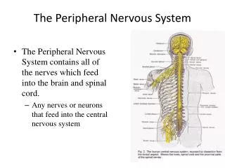
Basic Structural Components of the PNS • Sensory receptors – pick up stimuli from inside or outside the body • Motor endings – axon terminals of motor neurons • Innervate effectors (muscle fibers and glands) • Nerves and ganglia • Nerves – bundles of peripheral axons • Ganglia – clusters of peripheral neuronal cell bodies
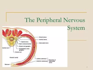
Basic Anatomical Scheme of the PNS in the Region of a Spinal Nerve Figure 14.2
Peripheral Sensory Receptors • Structures that pick up sensory stimuli • Initiate signals in sensory axons • Two main categories of sensory receptors • Free nerve endings of sensory neurons • Monitor general sensory information • Complete receptor cells – specialized epithelial cells or small neurons • Monitor most types of special sensory information
Peripheral Sensory Receptors • Sensory receptors also classified according to: • Location • Type of stimulus detected • Structure
Classification by Location • Exteroceptors – sensitive to stimuli arising from outside the body • Located at or near body surfaces • Include receptors for touch, pressure, pain, and temperature • Interoceptors – (visceroceptors) receive stimuli from internal viscera • Monitor a variety of stimuli • Proprioceptors – monitor degree of stretch • Located in musculoskeletal organs
Classification by Stimulus Detected • Mechanoreceptors – respond to mechanical forces • Thermoreceptors – respond to temperature changes • Chemoreceptors – respond to chemicals in solution • Photoreceptors – respond to light – located in the eye • Nociceptors – respond to harmful stimuli that result in pain
Classification by Structure • General sensory receptors • Widely distributed • Nerve endings of sensory neurons monitor: • Touch, pressure, vibration, stretch • Pain, temperature, proprioception • Divided into two groups • Free nerve endings • Encapsulated nerve endings
Free Nerve Endings • Abundant in epithelia and underlying connective tissue • Respond to pain and temperature • Monitor affective senses • Two specialized types of free nerve endings • Merkel discs – lie in the epidermis • Slowly adapting receptors for light touch • Hair follicle receptors – wrap around hair follicles • Rapidly adapting receptors
Unencapsulated Nerve Endings Table 14.1
Encapsulated Nerve Endings • Consist of one or more end fibers of sensory neurons • Enclosed in connective tissue • Mechanoreceptors • Include four main types
Encapsulated Nerve Endings • Meissner’s corpuscles • Spiraling nerve ending surrounded by Schwann cells • Occur in the dermal papillae • Rapidly adapting receptors for discriminative touch • Occur in sensitive, hairless areas of the skin
Meissner’s Corpuscles Table 14.1
Encapsulated Nerve Endings • Pacinian corpuscles • Single nerve ending surrounded by layers of flattened Schwann cells • Occur in the hypodermis • Sensitive to deep pressure – rapidly adapting receptors • Ruffini’s corpuscles • Located in the dermis and respond to pressure • Monitor continuous pressure on the skin – adapt slowly
Pacinian Corpuscles and Ruffini’s Corpuscles Table 14.1
Encapsulated Nerve Endings • Proprioceptors • Monitor stretch in locomotory organs • Three types of proprioceptors
Three Types of Proprioceptors • Muscle spindles – measure the changing length of a muscle • Imbedded in the perimysium between muscle fascicles • Golgi tendon organs – located near the muscle-tendon junction • Monitor tension within tendons • Joint kinesthetic receptors • Sensory nerve endings within the joint capsules
Proprioceptors Table 14.1
Structure of Proprioceptors Figure 14.4