Download
1 / 30
300 likes | 308 Views
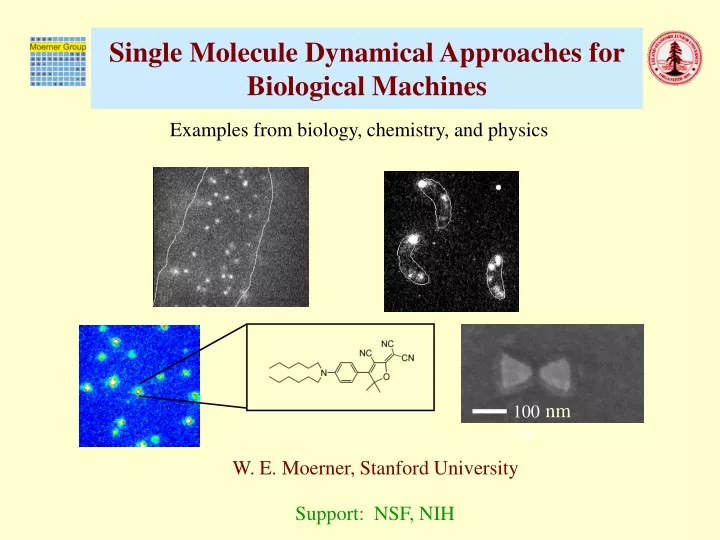
100 nm. nm. Single Molecule Dynamical Approaches for Biological Machines. Examples from biology, chemistry, and physics. W. E. Moerner, Stanford University. Support: NSF, NIH.

E N D
100 nm nm Single Molecule Dynamical Approaches for Biological Machines Examples from biology, chemistry, and physics W. E. Moerner, Stanford University Support: NSF, NIH
No ensemble averaging: direct observation of (static and dynamic) heterogeneity by measuring the full distribution Time-dependent state changes directly observable, without synchronization, and can detect rare intermediates Why Study Single Molecules? We can learn if the individual copies are “marching to different drummers”! This concept has the greatest potential in complex systems: crystals, amorphous materials, biological systems, and other condensed phases E. G., in biology, many processes occur at the single copy or few-copy level: DNA/gene expression, molecular motor operation, antigen recognition, protein synthesis, signaling, hormonal control, ...
1.1 nm Nanophotonics: Nanoscale Object + Light A Single Fluorescent Molecule as an Optical Nanoprobe • Typical single-molecule labels are only ~1 nm in size • Emission reports on local nanoenvironment (even though focal spot may be diffraction-limited) TMR Cy3 Terrylene Moerner, Dickson, and Norris, “Single-Molecule Nanophotonics in Solids,” Matls. Sci. and Engr. B48, 169 (1997).
P P O O O D F F F CCD OF O O D F F A CCD CCD SPAD SPAD (b) epi (a) TIR Microscope Configurations (c) confocal (d) NSOM
Optical Studies of Individual Moleculesphysical chemistry, chemical physics, biophysics, photonics www.stanford.edu/group/moerner Biophysics Enzymatic dynamics fpr beta-galactosidase Folding dynamics in chaperonins Dynamics, FRET: Fluorescent proteins GFP, cameleon, DsRed, BFP, ... Nanophotonics Single-photon light source Near-field optics Precision detection ... Diffusion: single molecules in cell membranes Polarization: kinesin motor orientations, dye molecules in polymers GroupOverview01.ppt
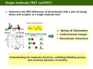
442nm 480nm CFP YFP M13 CaM +4 Ca2+ 442nm FRET 530nm 60 40 counts/bin 20 0 0 1 2 3 Time (sec) Detection of Local Calcium Concentrations by Fluorescence Resonance Energy Transfer From Single MoleculesSophie Brasselet, Erwin Peterman, Atsushi Miyawaki*, Roger Tsien*, and W.E. Moerner Dual-GFP “Cameleon” 2-color confocal microscopy single-molecule histograms SM time traces: D and A excess width at intermediate [Ca++] due to binding kinetics *Collaborators from UC San Diego See J. Phys. Chem B 104, 3676-3682 (2000).
Biological Example:Attaching a Bifunctional Label to Kinesin to Sense Local Orientation Hernando Sosa, Erwin Peterman, Larry Goldstein, WEM, Nat. Struct. Biol.8, 540-544 (June 2001) one view of expected label position another view of expected label position m Nucleotide ADP rhodamine ~80° - + microtubule starting structure: rat-brain kinesin of Sack et al. Biochem. 36, 16155 (1997) (Insight model)
ADP-Induced Rocking of the Kinesin Motor Domain Revealed by Single-Molecule Fluorescence Polarization Microscopy • Big Question: • How does kinesin move? • (processivity, coordination, … • Block, Vale, Yanagida, …) Hernando Sosa, Erwin Peterman, Larry Goldstein, WEM Nat. Struct. Biol. 8, 540-544 (June 2001) • Smaller Question: • What is the orientation of • a head relative to the • microtubule as a function • of nucleotide bound?
Single- and Multiple-Molecule Studies of Kinesin Motor Orientationon Microtubules Hernando Sosa, UCSD, now at Albert Einstein College of Medicine Erwin J. G. Peterman, UCSD, Stanford, now at Freie Univ. Amsterdam W. E. Moerner, UCSD, Stanford Larry S. B. Goldstein, HHMI and UCSD epifluorescence polarization microscopy, bi-functional attachment of fluorophore Nature Struct. Biol. 8, 540 (2001)
Single Molecules in Living Cells: A Challenging Regime • Key Issues: • Cellular autofluorescence – use long l’s, extreme care • What labeling strategy to use? • Approaches: • Extrinsic fluorophore bound to biomolecule, e.g., lipid, ligand, virus, etc. • Label is small, noninvasive, bright, but harder to target arbitrarily • Genetically encoded labels (autofluorescent proteins) • Powerful, but fusions large, possibly perturbative, emitters not as bright • Mixed approaches (FlAsH, enzymes, qdots, binding aptamers, many fluorophores on same biomolecule, …) Other workers: Schütz et al., Kubitschek, Yanagida, Braeuchle, Zhuang, Weiss…
Nonperturbative Sensing of Cell Membrane Dynamics Extracytoplasmic Region: Transmission image of the cell Fluorescence image of the cell (top plane) Fremont, et al. Science 1996, 272: 1001 Cy5 labeled peptide ligand MCC(95-103) GPI-linked native I-Ek Native I-Ek 5 mm D (mm2/s) Alberts et al, Molecular Biology of the Cell, Third edition, Garland Publishing, N.Y. 1994 Time lag (s) Vrljic, Nishimura, McConnell, Moerner Biophys. J. 2002, 2003 [Cholesterol] [Cholesterol]
Fremont, et al. Science 1996, 272: 1001 Real-time Images of Single MHCII Diffusion Cy5 100 ms/frame Vrljic, Nishimura, Brasselet, Moerner, McConnell, Biophysical Journal 83, 2681-2692 (2002)
Single-Molecule Dynamics Inside Living Caulobacter crescentus t = 1 s 1 μm 1 μm 1 μm Jason Deicha, Ellen M. Juddb,c, Lucy Shapirob, Harley H. McAdamsb, and W.E. Moernera aDepartment of Chemistry, Stanford University, Stanford, CA 94305-5080, USA bDepartment of Developmental Biology, Stanford University School of Medicine, Stanford, CA 94305, USA c Department of Applied Physics, Stanford University, Stanford, CA 94305, USA Work supported by: Deich, et al., PNAS 101, 15921 (2004).
Misfolded conformation Free Energy Native conformation X-ray Structures of GroEL & ES Reaction Coordinate GroEL Dye-tagged substrate (protein) GroEL Gro ES Gro EL (fixed) Tagging the substrate allows the observation of many turnovers w/o bleaching = unprecedented SM enzyme statistics 7 identical 57 kDa subunits form each ring 2 rings stack on top of each other to form full GroEL machine Binds to top of GroEL to provide isolated pocket for substrate proteins to fold in Assisted Protein Folding with the Chaperonin GroEL, ES SoYeon Kim, J. Frydman, W.E. Moerner Click-> Chaperones assist proteins out of kinetically trapped states and allow proteins to reach their most stable thermodynamic conformation
Single-Molecule Probes of Chaperonin-Assisted Protein Folding – A Status Report W. E. Moerner, So Yeon Kim, Stephan Hess Department of Chemistry in collaboration with Judith Frydman, Anne Meyer, Christoph Spiess Department of Biological Sciences Robert J. Twieg, Alexander Semyonov Kent State University Arthur Horwich, George W. Farr HHMI (Yale University Medical School) BioX-IIP Funding: NSF, BioX IIP, Alexander von Humboldt Stiftung BioX IIP Symposium, 31 July 2003
Outline • Introduction • -Molecular chaperones and the chaperonin GroEL/ES • -Key questions to be addressed – bulk and single-molecule • Nile red as a polarity-sensitive probe for GroEL/ES • -Bulk studies with nucleotide mimics • -Preliminary single-molecule experiments • FRET-sensed studies of assisted folding by GroEL/ES – development • of labeling protocol • (Use of Nile red to explore conformational changes in TRiC) • Summary
What is the exact sequence of binding/conformational changes driven by substrates and nucleotide? • Bulk: kinetics under controlled sequencing • Single-molecule: explore heterogeneity, follow time-dependence Cy3, Cy5-labeled Rhodanese GroES GroES ATP ATP Nile Red-labeled GroEL GroEL FRET Approaches to Explore Assisted Folding with GroEL/GroES What is the role of GroEL/ES – unfoldase? or a restricted geometry? • With a polarity sensitive fluorescent probe • With single-molecule FRET in the substrate, or between EL and substrate • What is the temporal relationship between conformational changes and substrate folding? • How many cycles are needed to fold?
Cys261 Apical domain Intermediate domain Equatorial domain ATP ATP ATP ATP 1. A GroEL Mutant with a Polarity Sensitive Probe Single-Cys mutant GroEL can be labeled with the a polarity sensitive fluorescent probe in the apical domain. Substrate • Why a polarity sensitive probe at this position? • The main interactions between GroEL and the substrate protein are hydrophobic – GroEL attracts substrates with exposed hydrophobic regions • The apical domain possesses binding sites for both the substrate and GroES. • ATP binding promotes the conformational change of the apical domain. • Thus, the binding/release of substrate, ATP, and GroES to GroEL should change the fluorescence of the probe molecule. GroES
less polar S2 Internal Conversion and Vibrational relaxation S1 Less polar solvent Solvent Relaxation More polar solvent hν ICT state hν hν S0 Photophysical Properties of Nile Red Nile red can be used as polarity sensitive probe due to its TICT (Twisted Intramolecular Charge Transfer) state. Nile red maleimide The fluorescence intensity of Nile red increases and the spectrum is blue-shifted as the solvent becomes more nonpolar.
Adding GroES Adding MDH Sample of Results from Bulk Study: GroEL-NR with unfolded substrate MDH (malate dehydrogenase) Adding Nucleotide Signal from GroEL-NR alone (1:1 NR:EL) • Following Kinetics of Binding Events and Conformational Changes: • +MDH, large change to nonpolar • +nucleotide or ES, more polar • Little dependence on order of adding substrate, ES, nucleotide • Some sequence-dependent effects with ADP-AlFx
10 mm 532 nm excitation Dichroic beamsplitter (545 nm long pass) 5 mm Microscope Tube lens 100 m pinhole 0 mm 0 mm 5 mm 10 mm Raman Notch 550 nm Long pass APD Confocal Imaging of Single Copies of GroEL-NR in Agarose Gels Experimental conditions: concentration ~10-9 M in 1.5% agarose gel excitation intensity ~2 kW/cm2 Confocal Setup 10 ms per point, 100100 points
Can We Use Lifetimes to Sense Polarity Changes? Uses basic relation: tF = FF x tRAD Bulk test shows little change in lifetime with addition of unfolded MDH! This implies that both the quantum yield and the radiative lifetime changed, or that there are two species, one fluorescent, and one nonfluorescent…
Cys 247 (buried, active site) Cys 254 (buried) Cys 263 (exposed) Cys 63 (exposed) Approach 2 : Cy3-Cy5 FRET on substrate protein • Use a mixture of Cy3 and Cy5 to label the two exposed cysteine residues of • the GroEL/ES substrate rhodanese. • Monitor events of substrate denaturation and refolding with GroEL/GroES by the • FRET efficiency. • Four different combinations of dyes in labeled protein will be; • Donor-Donor, Donor-Acceptor/Acceptor-Donor, and Acceptor-Acceptor J.H. Ploegman et al, J. Mol. Biol.127,149 (1979)
532 nm excitation Dichroic beamsplitter (reflect < 545 nm) 100 m pinhole Raman Notch 550 nm long pass Donor APD Dichroic beamsplitter (reflect < 630 nm) Acceptor APD 650-700 nm band pass Two-Color Confocal Microscopy of Single Molecules sample (Can also pump acceptor only)
Rhodanese labeled with Cy3 and Cy5, spincoated on cover slip, 10-9 M Acceptor Donor
Future Plans for Continuation of Collaboration • Publish results of bulk GroEL-NR study with MDH • Surface attachment of labeled GroEL • via His6 tag – easier to use with flow cell • Refinement of dual-labeling of rhodanese • FRET between labeled substrate and labeled EL • Alternate polarity-sensitive probe • Apply newly-developed assays to eukaryotic chaperonin TRiC
A New Class of Fluorophores for Single-Molecule Imaging • A bit of serendipity from separate research on photorefractive • polymers • Kallie Willets, Oksana Ostroverkhova, Meng He, • Robert J. Twieg, and W. E. Moerner • JACS online (10.1021/ja029100q, Jan. 2003) • Click->
100 nm Summary: Single Molecules as Nanoscale Probesin Biological Systems • A single molecule can be a reporter of local orientations, positions, proximity, E fields, polarity, dynamics, … for in vitro studies to in vivo work in cells • New fluorophores provide additional reporter functions in membranes and cells • Nanoscale metallic structures concentrate light and promise higher resolution, Raman scattering, and biological imaging