Download
1 / 62
650 likes | 805 Views
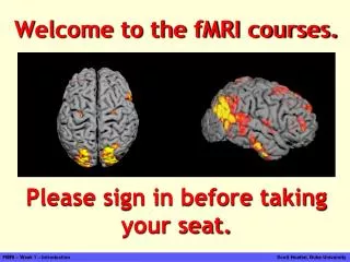
Welcome to the fMRI course. Please sign in before taking your seat. An Introduction to Functional MRI. FMRI Graduate Course (NBIO 381, PSY 362) Dr. Scott Huettel, Course Director. Some Introductions: People. Course Director:

E N D
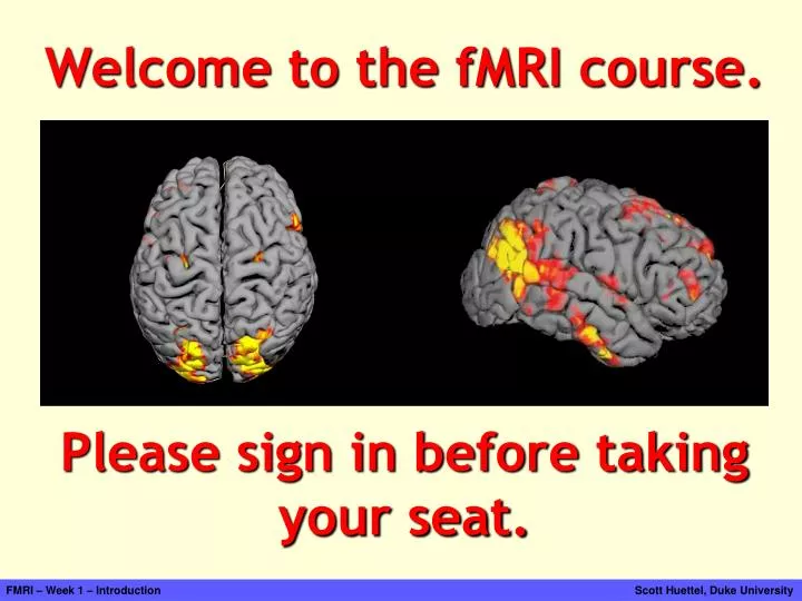
Welcome to the fMRI course. Please sign in before taking your seat. FMRI – Week 1 – Introduction Scott Huettel, Duke University
An Introduction to Functional MRI FMRI Graduate Course (NBIO 381, PSY 362) Dr. Scott Huettel, Course Director FMRI – Week 1 – Introduction Scott Huettel, Duke University
Some Introductions: People Course Director: Scott Huettel Associate Professor, Psychology & Neuroscience, BIAC, CCN Research Interests: Decision making, neuroeconomics Teaching Assistant: David V. Smith Graduate Student, Psychology & Neuroscience, IPCN Research Interests: Decision making, social rewards Jim Voyvodic Michele Diaz Allen Song FMRI – Week 1 – Introduction Scott Huettel, Duke University
Some Introductions: Places Duke Teaching and Learning Center, “The Link” Duke-UNC Brain Imaging & Analysis Center (BIAC) BIAC Offices and Analysis Laboratory, Bell Building www.biac.duke.edu MRI Scanners (3T, 4T), Duke Hospital FMRI – Week 1 – Introduction Scott Huettel, Duke University
Overview of the Course • Lectures • Wednesdays 3-4:30pm • Perkins -- “The Link”, Seminar Room 4 • Readings • Functional Magnetic Resonance Imaging (Huettel, Song, McCarthy) • Original papers, posted to website (optional) • Laboratories • Wednesdays 4:30-6:00pm • Additional times arranged with TAs and instructor (group) • David is planning on office hours on Tuesdays (in the Link) • Grading Basis • Attendance • Weekly laboratory exercises (group) • Self-assessment exercises • Mid-term examination • Project presentation (group) Course auditors are welcome to attend lectures! FMRI – Week 1 – Introduction Scott Huettel, Duke University
Course Textbook • First edition (2004): Required • Selected chapters from new edition (2008) will be provided by instructor • REQUIRED: Self-assessment questions available on accompanying CD FMRI – Week 1 – Introduction Scott Huettel, Duke University
Each week has lecture and laboratory components Labs start next week. In late September, you will form small groups for your fMRI projects. We’ll go over the project phase of the course in great detail around then. We will introduce the analysis package FSL in a session in this room. The midterm on 10/8. Auditors are welcome to take it for fun. There may be slight changes to the order and coverage of topics in the final weeks. Each group will present their projects at a session during the normal final examination period. FMRI – Week 1 – Introduction Scott Huettel, Duke University
Course logistics… or “What you need to do!” • Get a copy of the course textbook. • Find a partner for laboratory exercises. Most people will be in groups of 2; we can have one group of 3 as necessary. • Be aware of TA and computer availability for laboratories. • During the semester, download course materials from the class website: http://www.biac.duke.edu/education/courses/fall07/fmri/ (all materials will also be available on BlackBoard) FMRI – Week 1 – Introduction Scott Huettel, Duke University
Any questions? FMRI – Week 1 – Introduction Scott Huettel, Duke University
Outline for Today • Lecture: Introducing fMRI • What is fMRI? • History • Key concepts • Parts of a MR scanner • MR safety • Laboratory: Scanner Visit (Dr. Jim Voyvodic) • Scanner hardware • Stimulus presentation and recording hardware • Demonstration of real-time fMRI (??) Note: I will post all slides to the course web page! FMRI – Week 1 – Introduction Scott Huettel, Duke University
1. What is fMRI ? FMRI – Week 1 – Introduction Scott Huettel, Duke University
1. What is fMRI ? isn’t FMRI – Week 1 – Introduction Scott Huettel, Duke University
fMRI is not bumpology FMRI – Week 1 – Introduction Scott Huettel, Duke University
Phrenology claimed that bumps on the skull reflected exaggerated functions/traits • It lacked any mechanism underlying its claims. • It used anecdotal, rather than scientific, evidence. • Nevertheless, its central idea persisted: Localization of Function from Gall (c. 1810) Franz Joseph Gall (1758-1828) Johann Spurzheim (1776-1832) FMRI – Week 1 – Introduction Scott Huettel, Duke University
This is not a thought. This is not a thought. This is not an anti-thought. fMRI is not mind-reading FMRI – Week 1 – Introduction Scott Huettel, Duke University
fMRI is not a window on the brain “Mirror neuron activity in the right posterior inferior frontal gyrus – indicating identification and empathy - while watching the Disney/NFL ad.” rIFG “Ventral striatum activity – indicating reward processing - while watching the Disney/NFL ad.” ventStr [Citations omitted to protect the offenders.] FMRI – Week 1 – Introduction Scott Huettel, Duke University
fMRI is not invasive Positron Emission Tomography (PET) Intracranial Stimulation / Recording FMRI – Week 1 – Introduction Scott Huettel, Duke University
FMRI is… a technique for measuring metabolic correlates of neuronal activity • Uses a standard MRI scanner • Acquires a series of images (numbers) • Measures changes in blood oxygenation • Use non-invasive, non-ionizing radiation • Can be repeated many times; can be used for a wide range of subjects • Combines good spatial and reasonable temporal resolution FMRI – Week 1 – Introduction Scott Huettel, Duke University
Manipulation Techniques Lesions, TMS, Stimulation Measurement Techniques fMRI, PET, EEG fMRI is a Measurement Technique… BRAIN BEHAVIOR FMRI – Week 1 – Introduction Scott Huettel, Duke University
… that provides information about a wide range of topics. From what we see… (ocular dominance columns) … to what we feel. (the dread of an upcoming shock) Cheng, Waggoner, & Tanaka (2001) Neuron Berns et al. (2006) Science FMRI – Week 1 – Introduction Scott Huettel, Duke University
2. History of fMRI FMRI – Week 1 – Introduction Scott Huettel, Duke University
1920 1930 1940 1950 1960 1970 1980 1990 2000 Timeline of MR Imaging 1972 – Damadian patents idea for large NMR scanner to detect malignant tissue. 1924 - Pauli suggests that nuclear particles may have angular momentum (spin). 1937 – Rabi measures magnetic moment of nucleus. Coins “magnetic resonance”. 1985 – Insurance reimbursements for MRI exams begin. 1973 – Lauterbur publishesmethod for generating images using NMR gradients. MRI scanners become clinically prevalent. 1944 – Rabi wins Nobel prize in Physics. 1952 – Purcell and Bloch share Nobel prize in Physics. M R NMR becomes MRI 1973 – Mansfield independently publishes gradient approach to MR. 1990 – Ogawa and colleagues create functional images using endogenous, blood-oxygenation contrast. 1946 – Purcell shows that matter absorbs energy at a resonant frequency. 1959 – Singer measures blood flow using NMR (in mice). 1975 – Ernst develops 2D-Fourier transform for MR. 1946 – Bloch demonstrates that nuclear precession can be measured in detector coils. FMRI – Week 1 – Introduction Scott Huettel, Duke University
Early Uses of NMR • Most early NMR was used for chemical analysis • No medical applications • 1971 – Damadian publishes and patents idea for using NMR to distinguish healthy and malignant tissues • “Tumor detection by nuclear magnetic resonance”, Science • Proposes using differences in relaxation times • No image formation method proposed • 1973 – Lauterbur describes projection method for creating NMR images • Mansfield (1973) independently describes similar approach FMRI – Week 1 – Introduction Scott Huettel, Duke University
The First ZMR NMR Image Lauterbur, P.C. (1973). Image formation by induced local interaction: Examples employing nuclear magnetic resonance. Nature, 242, 190-191. FMRI – Week 1 – Introduction Scott Huettel, Duke University
Early Human MR Images (Damadian) FMRI – Week 1 – Introduction Scott Huettel, Duke University
Mink5 Image – Damadian (1977) FMRI – Week 1 – Introduction Scott Huettel, Duke University
Digression: 2003 Nobel Controversy Paul Lauterbur Peter Mansfield FMRI – Week 1 – Introduction Scott Huettel, Duke University
Raymond Damadian FMRI – Week 1 – Introduction Scott Huettel, Duke University
New York Times October 9, 2003 FMRI – Week 1 – Introduction Scott Huettel, Duke University
Nobel Press ReleaseOctober 6, 2003 Summary Imaging of human internal organs with exact and non-invasive methods is very important for medical diagnosis, treatment and follow-up. This year's Nobel Laureates in Physiology or Medicine have made seminal discoveries concerning the use of magnetic resonance to visualize different structures. These discoveries have led to the development of modern magnetic resonance imaging, MRI, which represents a breakthrough in medical diagnostics and research. … This year's Nobel Laureates in Physiology or Medicine are awarded for crucial achievements in the development of applications of medical importance. In the beginning of the 1970s, they made seminal discoveries concerning the development of the technique to visualize different structures. These findings provided the basis for the development of magnetic resonance into a useful imaging method. Paul Lauterbur discovered that introduction of gradients in the magnetic field made it possible to create two-dimensional images of structures that could not be visualized by other techniques. In 1973, he described how addition of gradient magnets to the main magnet made it possible to visualize a cross section of tubes with ordinary water surrounded by heavy water. No other imaging method can differentiate between ordinary and heavy water. Peter Mansfield utilized gradients in the magnetic field in order to more precisely show differences in the resonance. He showed how the detected signals rapidly and effectively could be analysed and transformed to an image. This was an essential step in order to obtain a practical method. Mansfield also showed how extremely rapid imaging could be achieved by very fast gradient variations (so called echo-planar scanning). This technique became useful in clinical practice a decade later. FMRI – Week 1 – Introduction Scott Huettel, Duke University
1920 1930 1940 1950 1960 1970 1980 1990 2000 Timeline of MR Imaging 1972 – Damadian patents idea for large NMR scanner to detect malignant tissue. 1924 - Pauli suggests that nuclear particles may have angular momentum (spin). 1937 – Rabi measures magnetic moment of nucleus. Coins “magnetic resonance”. 1985 – Insurance reimbursements for MRI exams begin. 1973 – Lauterbur publishesmethod for generating images using NMR gradients. MRI scanners become clinically prevalent. 1944 – Rabi wins Nobel prize in Physics. 1952 – Purcell and Bloch share Nobel prize in Physics. M R I f NMR becomes MRI 1973 – Mansfield independently publishes gradient approach to MR. 1990 – Ogawa and colleagues create functional images using endogenous, blood-oxygenation contrast. 1946 – Purcell shows that matter absorbs energy at a resonant frequency. 1959 – Singer measures blood flow using NMR (in mice). 1975 – Ernst develops 2D-Fourier transform for MR. 1946 – Bloch demonstrates that nuclear precession can be measured in detector coils. FMRI – Week 1 – Introduction Scott Huettel, Duke University
Physiology (BOLD Contrast) Blood-Oxygenation-Level Dependent contrast FMRI – Week 1 – Introduction Scott Huettel, Duke University
Using MRI to Study Brain Function Kwong, et al., 1992 Visual Cortex FMRI – Week 1 – Introduction Scott Huettel, Duke University
Growth in fMRI : Published Studies Medline search on “functional magnetic resonance”, “functional MRI”, and “fMRI”. Year 2004 = ~1500; Years 2005+ > 2000 … FMRI – Week 1 – Introduction Scott Huettel, Duke University
3. Key Concepts FMRI – Week 1 – Introduction Scott Huettel, Duke University
Key Concepts • Contrast • Spatial Resolution • Temporal Resolution • Functional Resolution FMRI – Week 1 – Introduction Scott Huettel, Duke University
Contrast: Conceptual Overview FMRI – Week 1 – Introduction Scott Huettel, Duke University
Contrast: Anatomical Contrast: 1) An intensity difference between quantities: “How much?” 2) The quantity being measured: “What?” Contrast-to-noise: The magnitude of the intensity difference between quantities divided by the variability in their measurements. FMRI – Week 1 – Introduction Scott Huettel, Duke University
Contrast: Functional Contrast-to-noise is critical for fMRI: How effectively can we decide whether a given brain region has property X or property Y? FMRI – Week 1 – Introduction Scott Huettel, Duke University
Spatial Resolution: Voxels Voxel: A small rectangular prism that is the basic sampling unit of fMRI. Typical anatomical voxel: (1.5mm)3. Typical functional voxel: (4mm)3. FMRI – Week 1 – Introduction Scott Huettel, Duke University
~8mm2 ~4mm2 ~2mm2 ~1.5mm2 ~1mm2 Spatial Resolution: Examples FMRI – Week 1 – Introduction Scott Huettel, Duke University
Temporal Resolution • Determining factors • Sampling rate, usually repetition time (TR) • Dependent variable, usually BOLD response • BOLD response is sluggish, taking 2-3 seconds to rise above baseline and 4-6 seconds to peak • Experimental design • Most FMRI studies have temporal resolution on the order of a few seconds • With specialized designs and data acquisition, this can be improved to ~100ms FMRI – Week 1 – Introduction Scott Huettel, Duke University
FMRI – Week 1 – Introduction Scott Huettel, Duke University
FMRI – Week 1 – Introduction Scott Huettel, Duke University
Functional Resolution The ability of a measurement technique to identify the relation between underlying neuronal activity and a cognitive or behavioral phenomenon. Functional resolution is limited both by the intrinsic properties of our brain measure and by our ability to manipulate the experimental design to allow variation in the phenomenon of interest. FMRI – Week 1 – Introduction Scott Huettel, Duke University
4. MRI Scanners FMRI – Week 1 – Introduction Scott Huettel, Duke University
GE 3T Scanner (cf. BIAC’s) FMRI – Week 1 – Introduction Scott Huettel, Duke University
FONAR 0.6T MR Operating Room Phillips 0.6T Open Scanner Phillips 3T Scanner (Vanderbilt) Siemens 3T Scanner FMRI – Week 1 – Introduction Scott Huettel, Duke University
Main Components of a Scanner • Magnetic: Static Magnetic Field Coils • Resonance: Radiofrequency Coil • Imaging: Gradient Field Coils • Shimming Coils • Data transfer and storage computers • Physiological monitoring, stimulus display, and behavioral recording hardware FMRI – Week 1 – Introduction Scott Huettel, Duke University
1. Magnetic: Static Field Coils The scanner contains large parallel coilings of wires. These generate the main magnetic field (B0), which gives the scanner its field strength (e.g., 3T). FMRI – Week 1 – Introduction Scott Huettel, Duke University