Download
1 / 84
840 likes | 843 Views
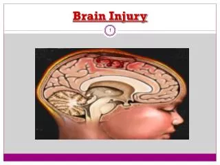
BRAIN INJURY BASICS. Karen Brewer, Ph.D. Clinical Neuropsychologist UT Southwestern Medical Center. Anatomy & Function of Major Brain Structures. Frontal Lobes Temporal Lobes Parietal Lobes Occipital Lobe Cerebellum Brain Stem Corpus Callosum Limbic System. Frontal Lobes.

E N D
BRAIN INJURY BASICS Karen Brewer, Ph.D. Clinical Neuropsychologist UT Southwestern Medical Center
Anatomy & Function of Major Brain Structures • Frontal Lobes • Temporal Lobes • Parietal Lobes • Occipital Lobe • Cerebellum • Brain Stem • Corpus Callosum • Limbic System
Frontal Lobes • Executive functions: • Planning • Organization • “Remembering to remember” • Self-monitoring • Higher-order thinking skills: • Problem solving • Reasoning • Cognitive flexibility • Sequencing
Frontal Lobes • Higher-order thinking skills: • Judgment • Insight • “Reading between the lines” • Attention/working memory • Motor functions – contralateral control • Basal area: • Bladder control • Taste/smell
Temporal Lobes • Memory: • Verbal memory – left • Visual memory – right • Auditory processing – both hemispheres • Language function (comprehension & expression) – usually left • 99% right-handers • 90-95% left-handers
Parietal Lobes • Tactile sensation - contralateral perception & integration of tactile information • Right-left discriminations • Left: reading comprehension, calculating • Right: visuospatial abilities (e.g., “mapping,” constructing)
Occipital Lobes • Vision • Color perception
Cerebellum • Motor control & coordination • Connects to brainstem
Corpus Callosum • Connects the two cerebral hemispheres • Made up of huge tracts of white matter that carry messages from one hemisphere to another
Limbic System • Amygdala – manages emotions (especially anger) • Hypothalamus – manages hunger & satiation • Thalamus – major relay station for sensory input & integration
Brain Stem • Passageway between the brain and the spinal cord • Basic life support functions: • Respiration • Heart rate/blood pressure • Where all the cranial nerves converge
TBI Basic Statistics • Annual incidence of head trauma in the United States - 2 million people per year • Incidence of head injury steadily increases until ages 15-25, then declines; it rises again after age 60+.
TBI Basic Statistics • 500,000 people will require hospitalization, and about 80,000 will suffer from some level of chronic cognitive and/or physical disability. • TBI is also the leading cause of death in adolescents and adults under 45 years of age, with an overall mortality rate of 25 per 100,000.
TBI Basic Statistics • Boys are twice as likely to suffer a brain injury as girls. • Men are injured twice as frequently as women, but die due to head injury four times more often.
TBI Basic Statistics • Brain Injury Severity Stats: • 76-85% are mild • 8-10% are moderate • 6-13% are severe
TBI Basic Statistics • Most common causes of head injury – all age groups: • Motor vehicle accidents (> 50%) • Falls (21%) • Violence (12%) • Sports/recreational injuries (10%).
TBI Basic Statistics • Health costs from TBI: estimated to be $35 billion per year in the U.S.
Classifying Brain Injury Penetrating/Open TBI • Open TBIs are characterized by the velocity and location of the missile at impact: • The higher the velocity of the missile, the more severe the injury. • The lower the path of the missile, the more severe the injury.
Classifying Brain Injury Closed TBI • Closed TBIs are classified as mild, moderate, or severe, depending on the neurological status of the patient soon after the injury
Classifying Brain Injury • Mild - GCS of 13–15, LOC of less than 30 minutes, and/or PTA of less than 1 hour • Moderate - GCS of 9–12, LOC of 1–24 hours, and/or PTA of 30 minutes to 24 hours • Severe - GCS of 8 or less, LOC of more 24 hours, and/or PTA of more than 1 day
Mild Traumatic Brain Injury Mild Closed TBI (aka “concussion,” minor brain injury) • Involves transient physiological disturbances • May cause trauma to the scalp and/or cervical spine, and, in some cases, contusions or hematomas • No obvious anatomic injury to the brain • Results from low-velocity head trauma and may involve transient loss of consciousness and/or memory of events immediately before and after trauma • Usually produces normal CT/MRI scans and neurologic assessments
Mild Traumatic Brain Injury • May result in post-concussion syndrome • Disabilities due to posttraumatic headaches, dizziness, sleep disturbances, and inability to concentrate and perform complex tasks • Over time, PCS may cause anxiety, depression, and/or other psychosocial problems
Moderate/Severe Traumatic Brain Injury • Associated with high velocity impact (e.g., motor vehicle accidents, assaults, & falls) • Diagnosed when there is any one of the following associated with a brain injury: • Contusion • Hematoma • Hydrocephalus • Skull fracture
Diffuse Axonal Injury • One of the most common and devastating types of brain injury (Iwata et al., 2004), occurring in almost half of all cases of severe head trauma. • Results from the motion of the brain within the skull, causing extensive damage to the axons (white matter). • This can produce a wide spectrum of injuries, ranging from brief physiological disruption to widespread axonal death.
Diffuse Axonal Injury • Secondary damage may occur after the initial injury: • Brain swelling • Cerebral edema/Increased intracranial pressure • Hypoxia • Hematoma/hemorrhage • Metabolic abnormalities • Hydrocephalus • Fat embolism • Excessive release of excitatory amino acids (e.g., glutamate overproduction increases hypoxic injury to the hippocampus) • Oxidative free-radical production • Disruption of neurotransmitters
Diffuse Axonal Injury • Delayed damage may occur as well: • White matter degeneration • Cerebral atrophy • Development of posttraumatic hydrocephalus • Development of posttraumatic seizures
QUIZ All things being equal (good pre-injury health, no post-injury complications, etc.), which of the following people is most likely to have the best outcome if he/she suffers a moderate brain injury? • 2-year-old • 22-year-old • 42-year-old
Common Cognitive Sequelae of TBI • Intellectual Decline • Verbal intelligence tends to be less vulnerable to mild and moderate brain injuries. • Nonverbal intellectual abilities (e.g., Performance IQ) more often affected due to problems with fluid problem solving and decreased information processing speed. • IQ tends to plateau 1-2 years after the injury, though improvements may be seen for up to 5 years.
Common Cognitive Sequelae of TBI • Attention – • Auditory & visual • Mediated primarily by the frontal lobes • Arousal • Simple attention (e.g., focusing on what someone is saying, ability to repeat numbers) • Sustained attention (maintaining focus for >5 minutes) • Selective attention (ignoring unimportant information while focusing on what is important) • Divided attention (attending to more than one thing at a time; driving)
Common Cognitive Sequelae of TBI • Information Processing - making sense of information presented to the brain • Mediated primarily by the frontal lobes and the white matter of brain • Quality vs. speed issues • Visual information processing • Auditory information processing • Working memory – requires manipulating information before processing it (e.g., multi-step mental math problems)
Common Cognitive Sequelae of TBI • Verbal Memory • Mediated by the left temporal lobe in >90% of people (>95% right-handers; >85% left-handers) • Learning (storage), retrieval, & recognition • Contextual information (e.g., stories, conversations) • Noncontextual information (e.g., lists, isolated facts) • Symbolic information (e.g., math equations, spelling)
Common Cognitive Sequelae of TBI • Visual Memory • Mediated by the right temporal lobe in most people • Learning, retrieval, & recognition • Contextual information (e.g., city streets, a famous person’s face) • Noncontextual information (e.g., an unfamiliar item)
Common Cognitive Sequelae of TBI • Reasoning/Problem-Solving • Mediated primarily by the frontal lobes • Common deficit in TBI, but often not recognized by the patient. • The area of function that often distinguishes the children & adults that adapt well post-injury from those who do not.
Common Cognitive Sequelae of TBI • Abstract Thinking/Planning/Organization • Mediated primarily by the frontal lobes, especially the prefrontal cortices • Often quite impaired in persons who are brain injured, but not recognized until after hospitalization.