Download
1 / 19
190 likes | 286 Views
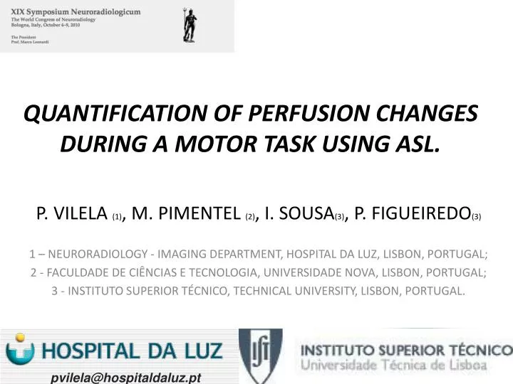
QUANTIFICATION OF PERFUSION CHANGES DURING A MOTOR TASK USING ASL. P. VILELA (1) , M. PIMENTEL (2) , I. SOUSA (3) , P. FIGUEIREDO (3) 1 – NEURORADIOLOGY - IMAGING DEPARTMENT, HOSPITAL DA LUZ, LISBON, PORTUGAL; 2 - FACULDADE DE CIÊNCIAS E TECNOLOGIA, UNIVERSIDADE NOVA, LISBON, PORTUGAL;

E N D
QUANTIFICATION OF PERFUSION CHANGES DURING A MOTOR TASK USING ASL. P. VILELA (1), M. PIMENTEL (2), I. SOUSA(3), P. FIGUEIREDO(3) 1 – NEURORADIOLOGY - IMAGING DEPARTMENT, HOSPITAL DA LUZ, LISBON, PORTUGAL; 2 - FACULDADE DE CIÊNCIAS E TECNOLOGIA, UNIVERSIDADE NOVA, LISBON, PORTUGAL; 3 - INSTITUTO SUPERIOR TÉCNICO, TECHNICAL UNIVERSITY, LISBON, PORTUGAL. pvilela@hospitaldaluz.pt
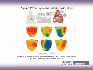
Objectives: Quantification of the CBF variation induced by the neural activity during a common motor task (finger tapping). rest CBF; activation CBF; Δ CBF; % Δ CBF CMRO2
Material & Methods Subjects:15 healthy volunteers (6F/9M, mean age 25.6) Stimulus: finger tapping by sequential thumb-digit opposition of the right hand Acquisition: 3.0T MRI system (Siemens Magnetom Verio) Paradigm Protocol: #1 ASL: 1 cycles rest/task (total acquisition time 3min51secX2). #2 BOLD-ASL: 5 cycles rest/task, 25 sec blocks (total acquisition time 4min12.5sec). 3.51 min 3.51 min 25 sec
Material & Methods ASL imaging: TR/TE = 2500ms/25ms 9 contiguous slices; 8mm slice thickness; 91x2 volumes, matrix 64x64; voxel size 3x3x8 mm3 (pulsed ASL sequence: PICORE Q2TIPS TI1 = 700 ms, TI1s = 1600 ms and TI2 = 1800 ms ) EPI BOLD-ASL imaging: TR/TE = 2500ms/11ms 9 contiguous slices; 8mm slice thickness; 101 volumes, matrix 64x64; voxel size 3x3x6 mm3 (pulsed ASL sequence: PICORE Q2TIPS TI1/TI2=700ms/1800ms) Subjects:15 healthy volunteers (6F/9M, mean age 25.6) Stimulus: finger tapping by sequential thumb-digit opposition of the right hand Acquisition: 3.0T MRI system (Siemens Magnetom Verio)
Analysis: • Cluster of activation: Z map Threshold: Z > 2.5 Cluster significance threshold: p < 0.05 Material & Methods Analysis: Standard General Linear Model (GLM) approach using FEAT from FSL [http://www.fmrib.ox.ac.uk/fsl] Pre-processing: motion correction, spatial smoothing FWHM = 5 mm, high-pass temporal filtering (f = 100ms) Protocol #1 Concatenation of the rest and activation scans in one single time-series data with the elimination of the first volume of each time-series, creating a single time-series dataset with 180 volumes (first 90 volumes – rest state and the other 90 volumes - activation state).
The respective CBF activation clusters given by statistical analysis were used to mask the respective quantitative maps, and the mean value within the cluster was calculated. Material & Methods Clusters of activation: CBF Quantification (ml / 100g / min) CBF rest; CBF activation Δ CBF = CBFact – CBFrest % Δ CBF = 100 x (CBFact – CBFrest) / CBFrest
The respective CBF activation clusters given by statistical analysis were used to mask the respective quantitative maps, and the mean value within the cluster was calculated. Material & Methods fractional BOLD signal change: ∆S/S0 (Davis et al. 1998). α = 0.38 (Grubb et al.1974; Mandeville et al. 1998). β = 1.3 (Bulte et al. 2009) M value= 4.3 (Chiarelli et al. 2007) Clusters of activation: CMRO2 Quantification
Results: Rest CBF Analysis Protocol #1 mean rest CBF: 61.0 ml/100g/min Protocol #2 mean rest CBF: 69.4 ml/100g/min Values Mean value
Results: Activation CBF Analysis Protocol #1 mean activation CBF: 104.8 ml /100g/min Protocol #2 mean activation CBF: 109.9 ml/100g/min Values Mean value
Results: Δ CBF Analysis Protocol #1 Δ CBF: 43.7 ml/100g/min Protocol #2 Δ CBF: 40.5 ml/100g/min Mean value Values
Protocol #1 %Δ CBF: 73±6 % • - Protocol #2 %Δ CBF:62±7 % Results: %Δ CBF Analysis Values Mean value
∆CBF = rCBFact – rCBFrest • Tissue type: p < 0.001; Correction: p = 0.031; Segmentation method: p < 0.001 GM Results: summary WM
fractional BOLD signal change: ∆S/S0 (Davis et al. 1998). α = 0.38 (Grubb et al.1974; Mandeville et al. 1998). β = 1.3 (Bulte et al. 2009) M value= 4.3 (Chiarelli et al. 2007) Results: CMRO2 Cerebral metabolic rate of oxygen (CMRO2). Evaluation Protocol #2 9 volunteers Mean %GM: 49% Mean %CBF: 62.49% (SE:8.52%) Mean BOLD SC: 0.71(SE:006%) Mean %CMRO2:22.56% (SE:5.48%) Aerobic (oxidative) metabolism
Perfusion /CMRO2 Coupling Results: CMRO2 CBF Cerebral metabolic rate of oxygen (CMRO2). Evaluation Protocol #2 9 volunteers Mean %GM: 49% Mean %CBF: 62.49% (SE:8.52%) Mean BOLD SC: 0.71(SE:006%) Mean %CMRO2:22.56% (SE:5.48%) CMRO2 / CBF: 0.33 (normal range 0.25-0.5) CMRO2
Conclusions These results show that both activation vs rest (protocol #1) and block design (protocol #2) functional protocols were capable to detect consistent variations in perfusion associated with a simple motor task. The block design has the advantages of requiring shorter acquisitions and allowing the acquisition of simultaneous BOLD contrast information, being the preferable approach for the evaluation of perfusion changes to endogenous stimuli.
Conclusions Perfusion ASL is a reliable method for the quantification (CBF) of the hemodynamic brain response to brain activation, and by combining the BOLD and CBF is able to estimate the cerebral metabolic rate of oxygen (CMRO2). This combined information (CBF & CMRO2) widens the scope of the ASL-fMRI applications as a non-invasive and reliable imaging approach to the study of the brain hemodynamic responses and metabolism activity.
Acknowledgments Authors: Marco Pimentel Inês Sousa Patrícia Figueiredo Technologists: Ana Cristina Santos Cidália Martins Fernando Gonçalves Ruben Teixeira pvilela@hospitaldaluz.pt
QUANTIFICATION OF PERFUSION CHANGES DURING A MOTOR TASK USING ASL. P. VILELA (1), M. PIMENTEL (2), I. SOUSA(3), P. FIGUEIREDO(3) 1 – NEURORADIOLOGY - IMAGING DEPARTMENT, HOSPITAL DA LUZ, LISBON, PORTUGAL; 2 - FACULDADE DE CIÊNCIAS E TECNOLOGIA, UNIVERSIDADE NOVA, LISBON, PORTUGAL; 3 - INSTITUTO SUPERIOR TÉCNICO, TECHNICAL UNIVERSITY, LISBON, PORTUGAL. pvilela@hospitaldaluz.pt