Download
1 / 50
620 likes | 1.32k Views
Cytogenetic Studies for Hematologic Malignancies in Korea. Introduction to Cytogenetic Studies External QA in Hematologic Malignancies Clinical Aspects of Molecular Cytogenetics: FISH, Array CGH. 혈액종양 관련 세포유전학 역사. Philadelphia chromosome (Ph) 1970 Banding technique

E N D
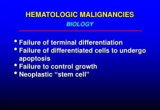
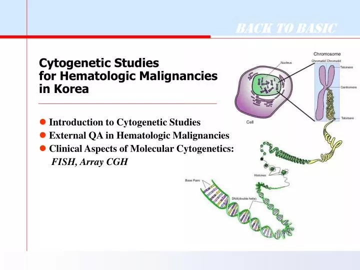
Cytogenetic Studies for Hematologic Malignancies in Korea • Introduction to Cytogenetic Studies • External QA in Hematologic Malignancies • Clinical Aspects of Molecular Cytogenetics: FISH, Array CGH
혈액종양 관련 세포유전학 역사 • Philadelphia chromosome (Ph) • 1970Banding technique • 1973 t(8;21)in AML-M2 • t(8;14)in Burkitt lymphoma/Leukemia • 1977 t(15;17)in AML-M3, t(4;11) • 1979 High-resolution banding technique • 1980 • 1982t(9;11) in AML-M5a, inv(3) in AML • 1983 inv(16) in AML-M4E • 1984 t(1;19) in ALL • t(1;3) in AML with dysmegakaryopoiesis • 1990 Molecular cytogenetics (FISH, CGH)
혈액종양 관련 세포유전학 역사 Cytogenetic & Gene Rearrangement • 1980 • 1983 MYC-IGH t(8;14) • 1984 ABL-BCR t(9;22), IGH-BCL2 t(14;18) • 1988 ATRA in APL Tx • 1990 • 1991 MLL t(4;11), E2A-PBX1 t(1;19) • PML-RARA t(15;17), AML1-ETO t(8;21) • DEK-CAN t(6;9) • 1993 CBFB-MYH11 inv(16), MLL-AF9 t(9;11) • 1997 TEL-AML1t(12;22)
KimSW, et al.: Banding patterns of normal human chromosomes. The Seoul Journal of Medicine 21(2):133-137(1980). Table 4. Karyotypic pattern in cases with CML 조혈기질환에 있어서 골수세포 염색체 분석에 관한 연구. 대한혈액학회지 23(2),1988
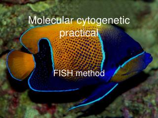
Giemsa Banding (G-Banding) Technique High-resolution Banding TechniqueUsing Methotrexate Cell Synchronization 1) PB/BM Culture RPMI 1640 FBS (15%) Pen-Strep L-Glutamine PHA PB/BM cells 3-5 hrs MTX(10-7M) 17 hrs 72 hrs Thymidine (10-5M) 2) Harvest 5 hrs 1) Colcemid (50 g/ml) treatment 2) Hypotonic Solution (KCl;0.075M) treatment 3) Fixation (methanol:Acetic acid=3:1) 3) Slide preparation & Staining (Giemsa-Trypsin) 4) Microscopy and Karyotyping 5) Printing (Photography)and Reporting
CCD camera Microscope Main Program Monitor PC Printer Computerized Image Analyzer Cytovision®
Conventional Cytogenetics in Hematologic Malignancies • 검사자 숙련도에 따라 슬라이드의 질적 차이가 많다 • 분열세포 적고, 염색체 길이 짧고, quality는 불량하다. • 복잡하고 다양한 핵형을 보이는 경우가 많다
ISCN 2005 • An International System for Human Cytogenetic Nomenclature (2005) • The complete citation for reference lists is: • ISCN (2005):An International System for Human Cytogenetic Nomenclature,Schaffer L.G., Tommerup N. (eds);S. Karger, Basel, 2005 ISCN (1985, 1991, 1995, 2005)
21. F. Mitelman, An International System for Human Cytogenetic Nomenclature (S. Karger, Basel, Switzerland, 1995).
Morphology (A) and karyotyping (B) of interspecies blastocysts derived from a human cord fibroblast transfer into enucleated bovine oocyte. An interspecies blastocyst at hatching were obtained 144 hours after culture and subsequently provided for chromosome analysis using a cytovision. Karyotyping shows 46 pairs of autosome and sex chromosome of XY. A.K. Tarkowski and J. Rossant, Haploid mouse blastocysts developed from bisected zygotes. Nature259 (1976), pp. 663–665
Philadelphia Chromosome (=Ph) der(22)t(9;22)(q34.1;q11.21) q11.2 BCR q34 ABL Ph (formerly Ph1) may be used in text, but not in the description of the karyotype, where der(22)t(9;22)(q34;q11.2) is recommended.
50,XX,+8,t(9;22)(q34;q11.2),+10,+19,+der(22)t(9;22)(q34.1;q11.21)50,XX,+8,t(9;22)(q34;q11.2),+10,+19,+der(22)t(9;22)(q34.1;q11.21) CML Blast Crisis - Karyotype
Practical Use of Cytogenetics in CML • Advantages ofCytogentics, compared to molecular DNA studiesfor BCR gene rearrangement • Distinguish betwn variant Ph and standard t(9;22) • Dectect other abnormalities : +8, i(17q),+Ph,+19 • Predict or confirm blast crisis • Give information regarding percentage of normal vs abnormal cells • Valuable after BMT to follow engraftment of the donor cells and identify possible relapse
AML-M2 with t(8;21)(q22;q22) • Usually AML-M2, occasionally M1 & M4 • Young individuals with good remission rate • Blasts containing a single thin Auer rod • RT-PCR : AML1/ETO fusion transcript
AML-M3 with t(15;17)(q22;q21) • The most specific clinical association in AML • Variants : t(15;Var;17), t(11;17), t(5;17) • RT-PCR : PML/RARA fusion transcript • Responsive to all trans-retinoic acid treatment 15 t(15;17) 17 RARA PML PML/RARA
AML-M4Eo with inv(16) • Young patients, organomegaly, abnormal eosinophils • Specifically associated with M4Eo in over 50% of cases • Favorable prognosis, High incidence of CNS relapse MYH11 CBFB CBFB-MYH11
Acute Myeloid Leukemias Proposed WHO Classification • AML with recurrent cytogenetic translocations • t(8;21), AML1(CBF)/ETO • t(15;17), PML/RAR • inv(16), CBF/MYH11 • 11q23(MLL) abnormalities • AML with multilineage dysplasia • AML and MDS, therapy related • AML not otherwise categorized
Normal Chromosome del 5q -7, del 7q del 9q del 20q +8 complex defects t(1;3), t(2;11) MDS t(3;3), inv(3) t(6;9), inv(16) t(8;21) t(9;22) t(v;11) t(15;17) AML
Myeloid Malignancy Secondary to Radiotherapy or Chemotherapy • Clinical features • AML-M1,M2,M6 • Lower remission rate and long-term reponse • Cytogenetics • Usually absence of t(8;21), t(15;17) & inv(16) • Usually complex karyotype • Common abnormalities: • -5, del(5q) in chemotherapy, • -7, del(7q) in radiation therapy, • Abnormalities of 3p, 11q23, 12p, and 17
Cytogenetics in Acute Lymphoblastic Leukemia • Poorly spread, short • Fuzzy chromosomes • Indistinct bands • Low success rate • 50% in conventional culture • 76% Clarkson (1985)
Ploidy Groups in Childhood ALL • Hyperdiploid >50 28% Early pre-B immunophenotype Found at the age of 2-10 years Lower WBC count, Favorable prognosis • Hyperdiploid 47-49 13% • Diploid 46 9% • Pseudodiploid 46 38% • Hypodiploid <46 6%
Lymphoid Neoplasm Proposed WHO Classification • B-Cell Neoplasm Precursor B-lymphoblastic leukemia/lymphoma • t(9;22), BCR/ABL • t(v;11), MLL rearranged • t(1;19), PBX/E2A • t(12;21), TEL/AML1 Mature B-cell neoplasm • T-Cell and NK-Cell Neoplasm • Hodgkin’s Lymphoma
Frequency of AML with specific chromosome defects 1)HallymUniversityMedicalCenter (1995) 2)UniversityofMinnesotaMedicalSchool
(External Proficiency Testing) 국내 유전검사 정확도 평가사업 ●법적 근거 • 생명윤리 및 안전에 관한 법률 (2005) • 유전자검사 시행기관 대상의 정확도 평가 사업을 시행 ●목적과 방법 • 유전검사의 정확도를 평가하고자 하는 목적 • 정도관리 물질을 각 검사기관에 발송하여 실제 검체와 같은 방법으로 검사한 결과를 상호 비교 ●대상기관 • 진료목적으로 유전자검사를 시행하는 모든 검사기관 • 순수 연구를 목적으로 하는 검사나, 그 시행기관은 제외
국내 세포유전검사 현황 • ●외부정도관리 사업: 검사의 정확도(숙련도) 평가, • External Quality Assurance (Proficiency Tests) • 대한임상검사정도관리협회(1997년) • 한국유전자검사평가원(2006년) (참고) 미국 CAP, 유럽 EMQN • ●국내 세포유전학검사실 • Conventional cytogenetics : 65 대학(종합)병원(43), 검사전문센터(8), 여성전문병원(13), 기타(1) • FISH 시행기관 : 22 BCR/ABL(19), AML1/ETO(19), TEL/AML1(15), MLL(15), X/Y(22)
세포유전검사 외부정도관리 사업 ●사업목표 • - 염색체 이상의 정확한 검출 • 정확한 명명법 준수 (ISCN 2005) ●사업내용 (10 cases/year) • - Metaphase 사진을 이용한 핵형분석 • 말초혈액, 양수, 골수 검체 등 • 핵형분석용 사진: 각 증례별 분열중기세포(metaphase) 5개씩 • 전혈 또는 골수 검체을 이용한 염색체검사 실시 ●증례선정: Cytogenetics Resource Committee
세포유전검사 결과 분석 ●평가기준: • - 참고기관의 80% 이상이거나, • 참여기관 다수의 의견일치(good or acceptable) • 외부정도관리위원회 검토 ●평가분석요소 • - M : modal chromosome number • S : sex chromosome designation • A : recognition of abnormalities • N : karyotype nomenclature (ISCN 2005) ●등급: Good / Acceptable // Unacceptable
세포유전검사 신빙도조사 결과 (1998) Constitutional Abnormalities Cancer Cytogenetics
세포유전검사 신빙도조사 결과 (2007) Constitutional abnormalities
세포유전검사 신빙도조사 결과 (2007) Cancer Cytogenetics
46,XX,t(9;22)(q34;q11.2)[2]/46,sl,inv(3)(q21q26.2)[3] • 46,XX,t(9;22)(q34;q11.2)[2]/46,sl,inv(3)(q21q26.2)[3] • 46,XX,t(9;22)(q34;q11.2)[2]/46,idem,inv(3)(q21q26.1)[3] • Stemline(sl) : the most basic clone of a tumor cell population listed first • Sidelines(sdl) : all additional deviating subclones, sdl1, sdl2, sdl3 • Idem : used only for a stemline with a single sideline
세포유전검사 정도관리사업 계획 • ISCN 2005 교육 프로그램 • 세포유전 검사방법 워크샵 • FISH 정도관리 시범사업 • 다양한 환자검체 확보 노력 • 표준시행지침서 주관기관: 삼성서울병원 유전검사실
WHO Classification “Real” disease entity No single ‘gold standard’ Morphology Immunophenotype Genetic features Molecular & cytogenetics Clinical feature
Chromosomal basis of Malignancy • Numerical abnormalities • polyploid : triploid(69,XXY), quadriploid(92,XXYY) • aneuploid : Trisomy, monosomy • Structural abnormalities • translocations • deletions • inversions • duplications etc. • Net loss of chromosomal material • inactivation of tumor suppressor genes • Net gain of chromosomal material • activation of protooncogenes • Relocation of sequences with no gain or loss of genetic material • new fusion genes : interfering regulatory control of genes
Molecular Cytogenetics (분자세포유전학) • Blurring the boundaries with cytogenetics and molecular biology • Bridging the gap between cytogenetic and molecular approaches • Based on fluorescent in situ hybridization (FISH) • Advances in FISH-based techniques • An important aim ⇒ Resolution↑ • The two crucial elements Target (resolution) Metaphase spreads (~5Mb) Interphase nuclei (50kb~2Mb) Chromatin strands using fiber FISH (5kb~500kb) DNA microarray (single-nucleotide level) Probe
The new cytogenetics: blurring the boundaries with molecular biology Exciting advances in fluorescence in situ hybridization and array-based techniques are changing the nature of cytogenetics, in both basic research and molecular diagnostics. Nature Review Genetics 6, 782-92 (Oct 2005) FISH probes for different applications
Advances in FISH-based techniques • Advances in metaphase spread analysis Multiplex-FISH (M-FISH) Spectral karyotyping (SKY)(Spectral karyotype imaging, SKI) Combined binary ratio labeling (COBRA) • Comparative genomic hybridization (CGH) on chromosomes • Applied to target metaphase chromosomes • Interphase cytogenetics
Conventional Copy Number Analysis Tools Spectral Karyotyping (SKY) Allows simultaneous visualization all the chromosomal pairs in different colors using chromosome specific probes More accurate than G-banding Karyotyping (G-banding) Provides a global view of metaphase chromosomal characteristics (number, type, shape etc) Each chromosome has a characteristic banding pattern that helps to identify them Fluorescent in situ Hybridization (FISH) More specific and sensitive than karyotyping Uses fluorescent probes to detect and localize the presence or absence of specific DNA sequences on chromosomes Resolution: 5 Mb – Metaphase 2 Mb – Interphase 0.5 Mb – Fibre FISH
Comparison of cytogenetic techniques for identifying chromosomal abnormalities.
Combining cytogenetic approaches to understand a complex chromosomal rearrangement Multiplex FISH Banding analysis Conventional CGH Array CGH Nat Rev Genet. 2005;6(10):782-92
Summary of Cytogenetics Technologies Karyotyping Provides a visual examination of the entire genome the best coverage but not the best resolution (≥ 5 Mb) Banding resolution differs from preparation to preparation FISH Sensitive (high resolution) Only provides information on tested regions, other aberrations will NOT be tested, i.e. not genome view Array-based Copy Number / CGH Analysis Single step global genome scan prevents FISHing expedition BAC (Bacterial Artificial Chromosome) Array Requires well characterized and high resolution clones High Resolution Oligonucleotide Microarray Highest resolution. Precise identification of gains and losses of genetic material.
FISH for hematologic malignancies *p16 signal 결손이나 중복 : 20 cases TEL/AML1 gene rearrangement : 14 cases MLL gene rearrangement : 5 cases BCR/ABL gene rearrangement : 4 cases Unpublished, SMC data
FISH profiles • ALL : MLL, BCR/ABL, ETV6/AML1, IgH, MYC, p16, E2A, chromosomes 4, 10, 17 • AML :ETO/AML1, PML/RARA, MLL, CBFB/MYH11, P53, Chromosomes 5, 7, 20 • CLPD : IGH, IGH/BCL2, IGH/CCND1, P53, MYB, ATM, 13q14.3, chromosome 12
Clinical considerations • Accuracy : detection of clonal abnormalities • Variations due to specimen quality, analysts, techniques • Supported by FISH, proficiency tests, periodic check of positive rate etc. • Turn-around time (TAT) • Technical TAT : 3-4 days • Ideal TAT : before final report of BM study • Practical TAT : 7~21 days • FISH tests • The more, the better? • Expecting Array CGH era