Download
1 / 45
530 likes | 843 Views
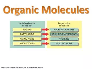
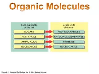
Organic Molecules. Six Functional Groups. Six Functional Groups. Early Origin-of-Life Experiments . Could the first steps of chemical evolution have occurred on ancient Earth?

E N D
Early Origin-of-Life Experiments Could the first steps of chemical evolution have occurred on ancient Earth? • To find out, Stanley Miller combined methane (CH4), ammonia (NH3), and hydrogen (H2) in a closed system with water, and applied heat and electricity as an energy source. • The products included hydrogen cyanide (HCN) and formaldehyde (H2CO), important precursors for more-complex organic molecules and amino acids. • In more recent experiments, amino acids and other organic molecules have been found to form easily under these conditions.
Nonpolar side chains Glycine (G) Gly Alanine (A) Ala Valine (V) Val Leucine (L) Leu Isoleucine (I) Ile No charged or electronegative atoms to form hydrogen bonds; not soluble in water Methionine (M) Met Phenylalanine (F) Phe Tryptophan (W) Trp Proline (P) Pro Polar side chains Figure 3-3 Partial charges can form hydrogen bonds; soluble in water Serine (S) Ser Threonine (T) Thr Cysteine (C) Cys Tyrosine (Y) Tyr Asparagine (N) Asn Glutamine (Q) Gln Acidic Basic Electrically charged side chains Charged side chains form hydrogen bonds; highly soluble in water Aspartate (D) Asp Glutamate (E) Glu Lysine (K) Lys Arginine (R) Arg Histidine (H) His
The Nature of Side Chains • The 21 amino acids differ only in the variable side chain or R-group attached to the central carbon • R-groups differ in their size, shape, reactivity, and interactions with water. (1) Nonpolar R-groups: Do not form hydrogen bonds; coalesce in water (2) Polar R-groups: Form hydrogen bonds; readily dissolve in water • Amino acids with hydroxyl, amino, carboxyl, or sulfhydryl functional groups in their side chains are more chemically reactive than those with side chains composed of only carbon and hydrogen atoms.
Primary Structure • A protein’s primary structure is its unique sequence of amino acids. • Because the amino acid R-groups affect a polypeptide’s properties and function, just a single amino acid change can radically alter protein function.
Normal amino acid sequence Single change in amino acid sequence 4 5 6 7 4 5 6 7 Figure 3-11 Normal red blood cells Sickled red blood cells
Secondary Structure • Secondarystructure results in part from hydrogen bonding between the carboxyl oxygen of one amino acid residue and the amino hydrogen of another. A polypeptide must bend to allow this hydrogen bonding—thus, -helices or -pleated sheets are formed. • Secondary structure depends on the primary structure—some amino acids are more likely to be involved in α-helices; while others, in β-pleated sheets. • Secondary Structure increases stability by way of the large number of hydrogen bonds.
Hydrogen bonds form between peptide chains. Hydrogen bonds Secondary structures of proteins result. -helix -pleated sheet Ribbon diagrams of secondary structure. Arrowheads are at the carboxyl end of the arrows -pleated sheet -helix
Tertiary Structure • The tertiarystructure of a polypeptide results from interactions between R-groups or between R-groups and the peptide backbone. These contacts cause the backbone to bend and fold, and contribute to the 3D shape of the polypeptide. • R-group interactions include hydrogen bonds, van der Waals interactions, covalent disulfide bonds, and ionic bonds. • Hydrogen bonds can form between hydrogen atoms and the carboxyl group in the peptide-bonded backbone, and between hydrogen atoms and atoms with partial negative charges in side chains.
Tertiary Structures of Proteins Interactions that determine the tertiary structure of proteins Hydrogen bond between side chain and carboxyl oxygen Ionic bond Hydrophobic interactions (van der Waals interactions) Hydrogen bond between two side chains Disulfide bond
Tertiary Structures of Proteins Tertiary structures are diverse. A tertiary structure composed mostly of -pleated sheets A tertiary structure rich in disulfide bonds A tertiary structure composed mostly of -helices
Van der Waals Interactions • van der Waals interactions are electrical interactions between hydrophobic side chains. Although these interactions are weak, the large number of van der Waals interactions in a polypeptide significantly increases stability. • Covalent disulfide bonds form between sulfur-containing R-groups. • Ionic bonds form between groups that have full and opposing charges.
Quaternary Structure • Some proteins contain several distinct polypeptide subunits that interact to form a single structure; the bonding of two or more subunits produces quaternarystructure. • The combined effects of primary, secondary, tertiary, and sometimes quaternary structure allow for amazing diversity in protein form and function.
Quaternary Structures of Proteins Cro protein, a dimer Hemoglobin, a tetramer
Simple Sugars • Monosaccharide • Single sugar • Ex. Glucose , Fructose • Disaccharide • Two sugars • Ex. Sucrose, Lactose, Maltose
Linear and Ring Forms Linear form of glucose Ring forms of glucose Oxygen from the5-carbon bonds to the1-carbon, resulting in a ring structure -Glucose -Glucose
Monosaccharides polymerize when hydroxyl groups react to form glycosidic linkages… Maltose (a disaccharide) -Glucose -Glucose Figure 5-4 The hydroxyl groups from the1-carbon and 4-carbon reactto produce an -1,4-glycosidiclinkage and water …between various carbons and with various geometries. Lactose (a disaccharide) -Galactose -Glucose In this case, the hydroxyl groups fromthe 1-carbon and 4-carbon react toproduct a -1,4-glycosidic linkage and water
Monosaccharides polymerize when hydroxyl groups react to form glycosidic linkages… Figure 5-4a -Glucose Maltose (a disaccharide) -Glucose The hydroxyl groups from the1-carbon and 4-carbon reactto produce an -1,4-glycosidiclinkage and water
…between various carbons and with various geometries. Figure 5-4b Lactose (a disaccharide) -Galactose -Glucose In this case, the hydroxyl groups fromthe 1-carbon and 4-carbon react toproduct a -1,4-glycosidic linkage and water
Polysaccharide • Complex Carbohydrate ( Starch) • Ex. Starch, Cellulose, Chitin • More than one ring structure
Cellulose, Chitin, Peptidoglycan Cellulose in plant cell wall Chitin in insect exoskeleton Peptidoglycan in bacterial cell wall
Glycoproteins: Identification Badge for Cells Glycoprotein Outside of cell Inside of cell
Lipids –Fats, Oils, Waxes, Steroids (cholesterol) • Unsaturated • Comes from plants and is liquid at room temperature • Ex. Corn oil, Olive oil, Sunflower oil • Better for you • Saturated • Comes from animals and is solid at room temperature • Ex. Bacon, animal fat • Bad for you
Nucleic Acids DNA • Deoxyribonucleic Acid • Phosphate, Deoxyribose sugar, Nitrogen Base • Double sided • Helical Structure • Found in nucleus RNA • Ribonucleic Acid • Phosphate, Ribose sugar, Nitrogen Base • Single sided • Can be various places in the cell depending on type
Only in RNA Only in DNA Nucleotide Nitrogen-containing bases Figure 4-1 Nitrogenous base Phosphate group Uracil (U) Pyrimidines Cytosine (C) Thymine (T) 5-carbon sugar Sugars Guanine (G) Adenine (A) Ribose Deoxyribose Purines
Figure 4-2 Condensation reaction Phosphodiester linkage
The sugar-phosphate spine of RNA The sequence of bases found in an RNA strandis written in the 5´ 3´direction Figure 4-3 Nitrogenousbases 3´ and 5´ carbonsjoined byphosphodiesterlinkage Unlinked 3´ carbon:New nucleotidesare added here
Hydrogen bonds form between G-C pairs and A-T pairs. Hydrogen bonds 5 3 Guanine Cytosine Figure 4-6b Sugar-phosphate backbone Adenine Thymine DNA contains thymine,whereas RNA contains uracil 5 3
Majorgroove Length of onecompleteturn of helix(10 rungs perturn) 3.4 nm Minorgroove Distancebetweenbases 0.34nm Width of thehelix 2.0 nm