Download
1 / 124
1.24k likes | 1.24k Views
To Review: Cells divide to stay small The cells in organisms that divide by mitosis are called somatic (body) cells The process of cell division is called mitosis Mitosis is a very small part of the cell’s life cycle Most of the cell’s life is spent in interphase

E N D
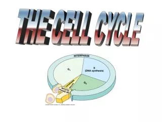
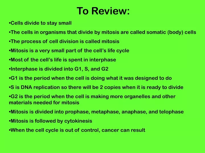
To Review: • Cells divide to stay small • The cells in organisms that divide by mitosis are called somatic (body) cells • The process of cell division is called mitosis • Mitosis is a very small part of the cell’s life cycle • Most of the cell’s life is spent in interphase • Interphase is divided into G1, S, and G2 • G1 is the period when the cell is doing what it was designed to do • S is DNA replication so there will be 2 copies when it is ready to divide • G2 is the period when the cell is making more organelles and other materials needed for mitosis • Mitosis is divided into prophase, metaphase, anaphase, and telophase • Mitosis is followed by cytokinesis • When the cell cycle is out of control, cancer can result
To Preview: • The reproductive structures in animals are called testes in males and ovaries in females • The cells in testes divide by meiosis to become sperm • The cells in ovaries divide by meiosis to become eggs • The cells in organisms that divide by meiosis are called gametes (sex cells; eggs and sperm are gametes) [remember that cells in an organism that divide by mitosis are called somatic cells] • Both mitosis and meiosis involve chromosomes • Scientists can analyze chromosomes using a karyotype • Sometimes meiosis or mitosis doesn’t go perfectly and chromosomal disorders can result • One type of chromosomal disorder that can result from an error in meiosis (or mitosis) is called a nondisjunction disorder
Differences Among Species • Each organism has a characteristic number of chromosomes • The number is constant with the species • Potatoes, plums, and chimpanzees all have 48 chromosomes
GENE • Units of information • Segment of DNA that codes for a protein • CHROMOSOME • Composed of DNA and protein called chromatin • Consist of 2 identical chromatidsattached by a centromere • Before a cell divides, chromatids separate to ensure that there is identical DNA in both cells
Somatic Cells • Any cell in the body other than a sperm or egg cell. Copied by mitosis • Have 46 chromosomes (23 pairs) – 23 of the chromosomes come from the mother and 23 chromosomes come from the father • These pairs of chromosomes are homologous. This means that each of the 23 chromosomes that came from the father has a corresponding chromosome from the mother. • A cell that contains both sets of homologous chromosomes is said to be diploid = two sets and is represented by the symbol 2N. In humans, the diploid number of chromosomes 46 [2N = 46]; 2N = 4 in fruit flies
Gametes – Sex Cells • Sperm or egg cell • Contain 1 set of chromosomes • 23 in humans; 2 in fruit flies • Said to be haploid (N); In humans N =23. Half as many as diploid • Fusion of 2 gametes = fertilization • Fertilized egg cell is called a zygote • 23 chromosomes + 23 chromosomes = 46 chromosomes (N + N = 2N) - Created by meiosis
CHROMOSOMES 1. Autosomes • 22 of the 23 pairs of chromosomes (44 of 46 chromosomes) – these are the body or somatic cells 2. Sex chromosomes • 1 of the 23 pairs (2 of the 46 chromosomes) • X and Y chromosomes in many organisms • In most organisms, if there is a Y it is male • XY in humans is male • If there is no Y, it is a female • XX is a normal female in humans • Who determines the sex of the organism? • XO – O refers to no chromosome • birds and butterflies have an XO system • In humans, XO is a genetic disorder called Turner’s Syndrome
This is a photograph of the chromosomes of a cell after DNA replication has occurred. This is typically when scientists try to photograph a squash of the nucleus, which hopefully results in the chromosomes being separated out. If a karyotype is going to be constructed, a computer can sort the chromosomes, line them up by size, and look for any abnormalities. It can (and used to be) be done “by hand”: the photograph taken through the microscope would be magnified and printed, then the chromosomes would be cut apart with scissors and sorted and ordered by hand.
Karyotype –magnified photograph of chromosomes grouped in order from largest to smallest in pairs; used to analyze if an individual has a missing or extra chromosome How many autosomes are shown? How many sex chromosomes? Is this from a male or a female? How do you know? This is a human karyotype. How many chromosomes are shown? 1 2 3 4 5 6 7 8 9 10 11 12 13 14 15 16 17 18 19 20 21 22
Karyotype • FISH: Florescent Imaging • Specific genes are dyed with radioactive elements causing them to be more easily detected in a karyotype
Part 2: Reproduction
Reproduction • Asexual– reproduction resulting from mitosis (or a similar process) that involves only one parent; the offspring are geneticallyidentical to the parent (a clone) • Sexual – reproduction resulting from an exchangeof genetic material; in most organisms this involves the joining or fusion of gametes (formed during meiosis)
Ways of Reproducing Asexually Binary Fission – separation of a parent into 2 or more individuals, ex. bacteria
Budding– a bud is formed and a new individual splits off from existing one. Ex. jellyfish, corals, yeast
Regeneration– renewal/regrowth of an organism from a part of that organism; ex. starfish
Asexual Reproduction Disadvantages: DNA varies little between organisms, which may make organisms NOT be able to adapt to a changing environment Benefits/Advantages: Produce many offspring in short period of time withoutusing energy to produce gametes or to find a mate Offspring are perfectly adapted to current environment, therefore often used in stable environments
Sexual Reproduction Disadvantages: Sexual reproduction uses a lot of energy in the development and maintenance of gametes, as well as a lot of biochemical resources. Benefits/Advantages: Frequent production of new combinations of genes which leads to genetic variation. This flexibility in the gene pool of a population helps insure the survival of a species, especially if there is rapid or sudden change in the environment.
What is Meiosis? • Form of cell division that produces sex cells (gametes) used in fertilization. • Process by which haploid (N) cells called gametes are produced from diploid (2N) cells • During mitosis, the number of chromosomes in each cell is cut in half through the separation of the homologous (same) chromosomes in a diploid cell
How is Meiosis Different? • There are 2 divisions in meiosis • Meiosis I and meiosis II • The result is 4 cells instead of 2 • In meiosis II, the DNA is not replicated again (No interphase) • The ending number of chromosomes is23 in humans (egg has 23 and sperm has 23). This is haploid (n).
Tetrad - Structure containing 4 chromatides that form during meiosis • Homologous chromosomes exchange portions of their chromatids in a process called crossing-over • Results in exchange of genes between homologous chromosomes and will produce new combinations of genetically different chromatids Crossing over in Prophase I – results in “new” chromosomes which leads to some genetic change
Oogenesis & Spermatogenesis • After meiosis the cells have a few more steps before they are eggs & sperm • Spermatogenesis: 4 sperms are created after the cell is modified to have flagella • Oogenesis: eggs need more cytoplasm and organelles so the cytoplasm of 3 of the cells is absorbed by 1. This results in 1 large egg cell and 3 polar bodies that get reabsorbed by the body.
Mitosis Division of somatic/body cells 1 division = 2 cells Daughter cells identical Diploid cells (2N) =46 Chromosome # identical to parent cell Used for growth and repair Meiosis Division of gametes (sex cells—eggs or sperm) 2 divisions = 4 cells Daughter cells different (crossing over; independent assortment) Haploid cells (N)=23 Chromosome # half of parent cell Mitosis Vs. Meiosis
How does meiosis promote genetic variation? • Independent Assortment - Each chromosome is randomly sorted independently of other chromosomes during metaphase I & II, so for a human who has 46 chromosomes, there are around 8 millionpossible combinationsfor a sperm or egg cell.(Imagine that the chromosomes are likecards being shuffled then dealt)
How does meiosis promote genetic variation? • Crossing Over - During prophase 1, remember that chromosomes come together in their homologous pairs forming tetrads. Sometimes, these pairs will swap the same segment of genes, forming a chromosome that has some of mom’s and some of dad’s DNA combined.
How does meiosis promote genetic variation? • Random Fertilization – the fertilization of an egg and sperm is completely random
Can you determine which children belong to which couple? Genetic variation is important in creating individuals! From Pearson Education
Answer: The first and last picture belong to couple 1. The middle pictures belong to couple 2.
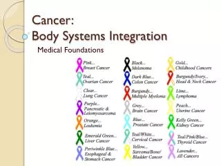
Part 4: Karyotypes, Nondisjunction,( & Chromosomal Disorders)
By looking at a karyotype, you should be able to: • Determine the sex/gender of the organism, • Determine if the organism is “normal” or has a chromosomal disorder, • Identify where a) the disorder is located b) what type of disorder (either nondisjunction or malformation of a chromosome) and possibly the specific name of the genetic disorder 4. Determine the number of autosomes and sex chromosomes present.
Humans have 23 pairs of chromosomes. • The last pair (#23) are the sex chromosomes. (XX-female, XY-male) • All others are called autosomes.
A karyotype is created from a picture of a cell during mitosis when the chromosomes are condensed and look like little Xs. Then a computer sorts the chromosomes into identical pairs and arranges them from largest to smallest (in length) and puts the 2 sex chromosomes at the end (pair 23). A doctor or other professional then analyzes the information shown for errors or abnormalities and uses them for an initial diagnosis.
Sometimes during meiosis when gametes are made, the chromosomes fail to separate correctly at anaphase I or anaphase II. This failure to separate is called nondisjunction. Results in the wrong number of chromosomes in a cell. Nondisjuction increases with age of females, or exposure to radiation and hazardous chemicals Most nondisjunction disorders are lethal to the developing organism. However, there are a few that do allow the organism to continue developing with varying degrees of effect. Nondisjunction could result in one of the zygotes (once egg & sperm unite) formed having only one copy of the affected chromosome. This is called monosomy. Another zygote could have 3 copies of one chromosome. This is called trisomy.
Common Nondisjunction Disorders Klinefelter's Syndrome: Trisomy+ of sex chromosomes • One or more extra sex chromosomes (i.e., XXY or XXXY); • 1 out of every 500-1,000 newborn males • Presence of Y chromosome directs development into a male • Underdevelopment of testes; usually infertile; taller than average; less hair than average • Although some lower scores on standardized tests have been reported, this is not necessarily the case. • Can be treatable with hormones
23 Klinefelter’s Karyotype Look at Chromosomes #23
Common Nondisjunction Disorders Turner’s Syndrome: Monosomy of the sex chromosomes • One sex chromosome, XO • 1 in 2500 female births • Absence of Y chromosome develops into female • Abnormal development of ovaries and secondary sexual characteristics during puberty; often infertile; shorter than normal • “Webbing" of the skin of the neck (extra folds of skin extending from the tops of the shoulders to the sides of the neck)
Common Nondisjunction Disorders Down Syndrome: Autosomal disorder • Trisomy 21, most common birth defect • 1 per 800 to 1,000 births (risk goes up as mother’s age goes up) • Mild to moderate learning disabilities (sometimes severe) • Eyes that slant upward and small ears that may fold over slightly at the top. Their mouth may be small, making the tongue appear large. Their nose also may be small, with a flattened nasal bridge
Down Syndrome Karyotype • Where did nondisjunction occur? (circle it in the karyotype) • What is the sex of this person? ________
Common Nondisjunction Disorders Patau Syndrome: Autosomal disorder, • Trisomy 13, rarely live past infancy • Extra fingers or toes (polydactyly) • Deformed feet, known as rocker-bottom feet • Neurological problems such as small head (microcephaly), failure of the brain to divide into halves during gestation (holoprosencephaly), severe mental deficiency • Facial defects such as small eyes (microphthalmia), absent or malformed nose, cleft lip and/or cleft palate • Heart defects (80% of individuals) • Kidney defects