Download
1 / 10
100 likes | 122 Views
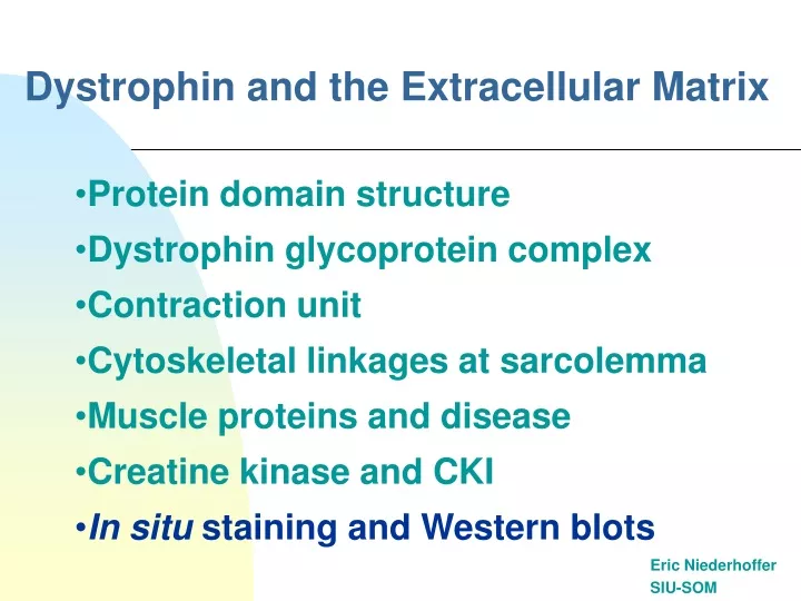
Dystrophin and the Extracellular Matrix. Eric Niederhoffer SIU-SOM. Protein domain structure Dystrophin glycoprotein complex Contraction unit Cytoskeletal linkages at sarcolemma Muscle proteins and disease Creatine kinase and CKI In situ staining and Western blots.

E N D
Dystrophin and the Extracellular Matrix Eric Niederhoffer SIU-SOM • Protein domain structure • Dystrophin glycoprotein complex • Contraction unit • Cytoskeletal linkages at sarcolemma • Muscle proteins and disease • Creatine kinase and CKI • In situ staining and Western blots
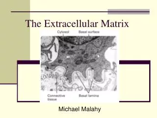
Dystrophin Domain Structure Domains of the dystrophin molecule. The N terminus contains the primary actin-binding site, whereas the C terminus contains the -dystroglycan, dystrobrevin, and syntrophin-binding sites. The N- and C-terminal domains are connected by 24 spectrin-like repeats, some of which have been shown to bind actin. The four "hinge" regions are denoted H1-H4. http://www.pnas.org/cgi/content/full/97/25/13464/F2
Dystrophin Glycoprotein Complex Schematic representation of the dystrophin-associated glycoprotein complex. The N-terminal, actin-binding domain of dystrophin in purple is associated with the cortical actin. The C-terminal domain associates with b-dystroglycan and with a- and b-syntrophin and dystrobrevin. nNOS is known to interact with the syntrophins as well as with caveolin. http://www.pnas.org/cgi/content/full/97/25/13464/F1
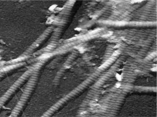
Contraction Units http://cellbio.annualreviews.org/cgi/content/full/18/1/637
Cytoskeletal Linkages at Sarcolemma http://cellbio.annualreviews.org/cgi/content/full/18/1/637
Muscle Proteins and Diseases http://www-ermm.cbcu.cam.ac.uk/0200488Xh.htm
contents spill out detected in blood Creatine Kinase and CKI • CKtotal 0-220 U/L CKMB 0-5 ng/mL • Muscle damage CKtotal elevated • CK index (CKI) = CKMB/CKtotal (x 100 for %) • Skeletal origin CKI <0.03 (3%) or use CKMM • Cardiac origin CKI >0.06 (6%) • If 0.03 (3%) > CKI <0.05 (5%) follow with Tn biomarkers Cell damage
In Situ Dystrophin • Normal dystrophin staining around the rim of muscle fibers Absent dystrophin: Duchenne muscular dystrophy • Left: No staining around the rim of muscle fibers. • Right: No staining of most muscle fibers. One "revertant" fiber with dystrophin staining. http://www.neuro.wustl.edu/neuromuscular/pathol/dmdpath.htm
Western Blots • Lane 3: Normal; Dystrophin has normal size and amount. • Lane 1: Becker dystrophy; Dystrophin has reduced abundance but normal size. • Lane 2: Becker dystrophy; Dystrophin has reduced size and abundance. • Lane 4: Duchenne dystrophy; Almost no protein is present. • Lane 5: Duchenne outlier; Dystrophin has severely reduced abundance. Western blot of dystrophin from dystrophinopathies. http://www.neuro.wustl.edu/neuromuscular/pathol/dmdpath.htm
Review Questions • What is dystrophin (domains, characteristics)? • Where is dystrophin found (interactions, role)? • How do you assess muscle damage? • How do you assess dystrophin levels?