Download
1 / 8
90 likes | 319 Views
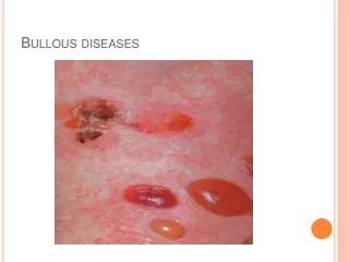
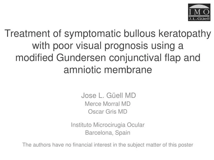
Treatment of symptomatic bullous keratopathy with poor visual prognosis using a modified Gundersen conjunctival flap and amniotic membrane . Jose L. Güell MD Merce Morral MD Oscar Gris MD Instituto Microcirugia Ocular Barcelona, Spain

E N D
Treatment of symptomatic bullous keratopathy with poor visual prognosis using a modified Gundersen conjunctival flap and amniotic membrane Jose L. Güell MD Merce Morral MD Oscar Gris MD Instituto Microcirugia Ocular Barcelona, Spain The authors have no financial interest in the subject matter of this poster
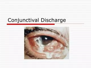
Purpose • To describe the use of a modified Gundersen conjunctival flap combined with amniotic membrane (AM) graft implantation to treat symptomatic bullous keratopathy (BK) in 5 eyes of 5 patients with poor visual prognosis.
Methods: Surgical technique (video file attached) 1 1. 360º conjunctival peritomy 2mm from the limbus
Methods: Surgical technique 2 2. Deepithelialization of the decompensated cornea preserving, if healthy, the limbal conjunctiva where the stem cell niches are located 3 3. Graft of AM sutured epithelial side up using a running 10-0 nylon suture at the periphery of the cornea with the knot buried in the corneal stroma. Attach the edges of the AM and the border of the peritomized conjunctiva using single 9/0 vycril sutures. The conjunctival border should lie over the AM. The AM graft covers the whole decompensated cornea and provides a basement membrane for conjunctival cells to grow on. Epithelialization occurs over the AM, which remains trapped until reabsortion is completed (Observe the section figure).
Methods: • Bandage contact lens • Topical steroids and antibiotics qid for three weeks, and tapered until complete reabsortion of the AM. • Outcome measures: • Resolution of the pain • Presence of ocular surface inflammation • Reinterventions
Results • 5/5 (100%) eyes - Immediate resolution of the pain and minimal postoperative inflammation. • 5/5 (100%) eyes - Epithelialization occurred over the AM. • 1/5 (20%) eye - The AM was reabsorbed before complete conjunctival epithelialization. Conjunctival reepithelialization over the central cornea was delayed. • No reinterventions required. • All the eyes asymptomatic for at least 16 months.
Case report Recurrent epithelial defects in a patient with post penetrating keratoplasty BK. Modified Gundersen conjunctival flap with amniotic membrane grafting was performed. Twelve months postoperatively, conjunctival vascular epithelium covers the cornea completely, providing sustained relief of the symptoms
Conclusion • The combination of a modified Gundersen conjunctival flap and a graft of amniotic membrane provided sustained relief of symptomatic bullous keratopathy • It is essential to implant the AM as a graft. The AM as a patch has only a temporary effect on relieving the pain • With this technique, conjunctival manipulation and anatomical distortion are significantly reduced: • The conjunctiva is not pulled over the cornea –Therefore, the fundus of the conjunctival sac is not shortened • The conjunctival peritomy is circular and the conjunctiva is not pulled – Therefore, there is no distortion of the interpalpebral bulbar conjunctiva