Download
1 / 46
540 likes | 804 Views
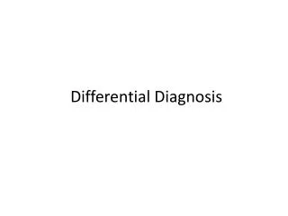
CPET in Differential Diagnosis. Gaye ULUBAY, MD, Assoc. Prof. Baskent University Faculty of Medicine Department of Pulmonary Diseases Ankara, Turkey. Pulmonary circulation. Peripheral circulation. VCO 2. CO2 PROD. EXPIRED. O2 FLOW. Creat PO 4. Muscle. Lungs. Heart and blood.

E N D
CPET in Differential Diagnosis Gaye ULUBAY, MD, Assoc. Prof. Baskent University Faculty of Medicine Department of Pulmonary Diseases Ankara, Turkey
Pulmonary circulation Peripheral circulation VCO2 CO2 PROD EXPIRED O2 FLOW CreatPO4 Muscle Lungs Heart and blood Mitochon INSPIRED PyrLac O2 CONSUM CO2 FLOW VO2 ENERGY • Obstructive • Restrictive • Infiltrative • Chest wall • CAD • Heart failure • Other HD • Anemia • Obesity • Myopathy • Detraining • Occlusive • Autonomic • Dysfunction • PAH • Thromboembolic • 1o & 2oPVD Wasserman K, Hansen FE Principles of Exercise Testing and Interpretation 2005
Determinants of VO2max Atmosphere Lungs:VE,V/Q, Diffusion Heart: Qt, SV, HR, BP Blood (Hb) Skeletal muscle (pump) Peripheral circulation: flow, capillary density, diffusion Muscle: fiber type, mass, mitochondria, O2 extraction, metabolism Johnson & Weisman.Clinical exercise testing, 2003
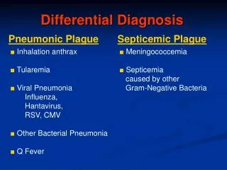
Why perform CPET ? • To demonstrate exercise limitation • To define cause of exercise limitation • To evaluate preoperatively • To determine morbidity and mortality • To follow up • To evaluate response to treatment
Dyspnea History, physical examination, CBC, Biochemical analysis, chest radiograph, ECG Abnormal; consider spesific diagnostic testing PFT, DLCO, ABG Results are normal BPT or cardiac tests Abnormal Normal Consider spesific diagnostic testing or treatment CPET Abnormal; Consider spesific diagnostic testing, treatment, and follow up normal; close follow-up Zeballos & Weisman.Clinical exercise testing, 2002
CPET parameters • Metabolic VO2, VCO2, R, AT, lactate • Cardiac HR, HRR, ECG, BP, O2 Pulse • Respiratory VE, TV, f, PETO2, PETCO2 • Gas exchange SpO2, VE/VCO2, VE/VO2 • Acid-base pH, PaO2, PaCO2 • Symptom Dyspnea, angina, leg fatique
CPET may fail in the evaluation of early/ mild disease as well as combined diseases
Would, but couldn’t Could, but didn’t
Is there an evidence suggesting a significant involvement of systems (respiratory, cardiovascular, muscle)? ERS Task force Eur Respir J 2007 Wasserman K, Hansen FE Principles of Exercise Testing and Interpretation 2005
Is Peak VO2reduced? Any disorder decreasing exercise capacity Peak VO2 (Panel 3)
Is PeakVO2at rest and exercise? Obesity Peak VO2, VO2-WR (Panel 3)
Is exercise limited by impaired O2 pulse? Heart diseases, peripheral/ PVH anemia, hipoxemia AT,VO2/WR, VCO2/WR,VO2/HR, VE/VCO2 (Panel 2,3,5,6)
Is exercise limited by reduced ventilatory capacity? Lung and chest wall diseases BR,Ventilatory response (Panel 1,7,9)
Is there a V/Q abnormality? PVD, Heart failure, Lung disease VD/VT,VE/VCO2-AT PETCO2 (Panel 4,6,9)
Poor effort or detraining ? Poor effort or detraining HRR, BR Peak R < 1.0, AT-N Panel (2,5,7,8)
Cardiovascular diseases; Heart F, PAH, CAD • Pulmonary diseases ; COPD, ILD • Obesity • Detraining
LEFT VENTRICULAR DYSFUNCTION VA/Q mismatching Blood (O2) flow H+La- CO2 • ATP production VD/VT pH Ventilatory requirement Inadequate myocardial contractility Dyspnea Dyspnea, fatique EXERCISE LIMITATION
Heart Failure • VD/VT- • HRR– N/ • WR • BR -N • VT - • VE –early increase • PECO2/PETCO2 • Peak VO2 • O2 pulse • VE/VCO2 • HR-VO2 is nonlineer • VO2/WR • AT/ VO2 • Early metabolic acidosis
Heart Failure • 20 Y, M • Dyspnea at rest and exercise • Nonsmoker • FEV1/FVC= 75 % • FEV1= 70 % • ECHO; EF= 25 %
COPD ± PULMONARY VASCULARDISEASE Work of breathing V/Q mismatching Blood flow ATP PaO2 VD/VT Lactate FEV1 and elastic recoil pH VCO2 Ventilatory recruitment Muscle fatigue Myopathy Ventilatory capacity + EXERCISE LIMITATION
COPD • VD/VT- • HRR– N/ • VT - • WR • VE - • PECO2/PETCO2 • Peak VO2 • O2 pulse • VE/VCO2 –AT • BR - • VO2/WR • AT/ VO2
Normal VO2 COPD Heart diseases W.R. REST EXERCISE
COPD • 62 Y, M • History of 50 pack-years cigarette smoking • Diagnosis of COPD • FEV1= 0.99L • FEV1/FVC= 60 % • MVV= 44L/min • VE= 42L/min
COPD Control COPD Control • 43 stable patients with COPD and 14 healthy controls • PFTs and CPET were performed • Hyperinflation determined at peak exercise, even no findings at rest Ulubay G, Görek A, Savaş S,Öner Eyüboğlu F. Tuberk Toraks, 2005
COPD ± PULMONARY VASCULARDISEASE Work of breathing V/Q mismatching Blood flow ATP PaO2 VD/VT Lactate FEV1 and elastic recoil pH VCO2 Ventilatory recruitment Muscle fatigue Myopathy Ventilatory capacity + EXERCISE LIMITATION
PAH • Peak VO2 • VE - • O2 pulse • VE/VCO2 –AT • VE/VCO2 • VD/VT- • AT VO2 • PETCO2 • PETO2 • BR – N • VT - • PECO2/PETCO2 • VO2/WR
PETCO2 PECO2, PECO2/PETCO2 Secondary to increased VD/VT PECO2/PETCO2 Ratio: 4 Groups PECO2: 4 Groups PETCO2: 4 Groups 0.80 35 50 0.75 45 30 0.70 Normal Normal Normal 40 0.65 COPD COPD COPD Ratio 35 25 0.60 HF PETCO2, mm Hg PECO2, mm Hg HF HF 0.55 30 PAH PAH PAH 20 0.50 25 0.45 20 15 0.40 Rest Unloaded AT Peak Rest Unloaded AT Peak Rest Unloaded AT Peak Activity Activity Activity n: 25 COPD, n: 25 LVF, n: 25 PVD patients Hansen JE, Ulubay G, Chow BF, Sun XG, Wasserman K. Chest 2007
73Y, F Complaint of dyspnea 1998- diagnosis of CREST syndr 2003- RT plus Tamoxifen for breast Ca Nonsmoker 2003- ECHO; PAP= 45mmHg Cpx= Bilateral crackles DLCO= 54 %, DLCO/VA= 58 % MVV= 66 L/min VO2/WR= 5.3 f=54/min Desaturated on test PHT
INTERSTITIAL LUNG DISEASES Parenchymal destruction /scarring Capillary destruction Pulmonary vascular resistance Hipoxemia VD/VT Elastic recoil Left ventricular filling Work of breathing Ventilatory recruitment Increased CO requirement Cardiac function Ventilatory impairment Circulatory impairment EXERCISE LIMITATION
ILD • Peak VO2 • O2 pulse • VE/VCO2 –AT • VE/VCO2 • WR • VO2/WR • AT/ VO2 • VD/VT- • R- (50/min) • HRR– / • BR -
TY 45 F • 45 Y, F • HRCT= diffuse bilateral parenchymal infiltrates • ECG=Normal • FVC =61% • TLC=84% • FEV1=58% • FEV1/FVC= 78 % • MVV = 56 L/min • DLCO = 18 % • f = 65/min • VD/VT 0.31 at rest 0.38 peak exercise
Obesity • High O2 cost • Peak VO2 is normal • Peak VO2/ body weight is low • Peak VO2/ height is normal • Normal VD/VT • Low PaO2 at rest that normalizes during exercise
Higher O2 cost than normal at rest and during exercise Obesity Normal VO2 W.R. REST EXERCISE Start exercise Wasserman K, Hansen JE Principles of Exercise Testing and Interpretation. 2005
45 Y, M • Complaint of exercise limitation • ECG=Normal • FEV1=65% • FEV1/FVC= 78 % • MVV = 140L/min • PVO2 = 126 % • ATVO2 = 70 %
Poor effort/ Detraining • Peak VO2 • HRR • AT determination rare • AT/ VO2 –N • Peak exercise R< 1.0 • WR • VO2/WR • BR – N • Caotic breathing pattern
37Y, M • Complaint of exercise limitation • ECG=Normal • FEV1=105% • FEV1/FVC= 78 % • MVV = 170L/min • PVO2 = 50 % • ATVO2 = undetermined
Normal Pretransplantation Posttransplantation Number of subjects 14 7 7 Age; mean in years (SD) 44 14 44 14 43 14 Sex Male 10 4 5 Female 4 3 2 Body mass index (kg/m2) 24 2 23 3 22 2 Hemoglobin (g/dL) 14 2 15 2 13 1 FEV1 (L/s) 3.12 1.04 4.81 1.09 3.08 0.61 FEV1/FVC (%) 80 6 88 4 78 5 Postoperative duration (mo) - - 19 7 LVEF, mean (SD) - 32 17 57 2 Peak VO2 (L/min) 2,24 0.64 1.11 0.36 1.45 0.33 HR (min) 160 15 120 25 114 41 MVV-VE max (L/min) 14 18 24 VE (L/min) 42 10 40 10 36 12 O2 pulse (mL/beat) 13.3 3 8.2 2.1 10.6 5.4 AT D etermined 14 2 - Undetermined - 5 7 FACTORS AFFECTING EXERCISE CAPACITY IN HEART TRANSPLANTATION RECIPIENTS Ulubay G. Ulasli SS. Sezgin A, Haberal M Clin Transplantation 2007
Ulubay G. Ulasli SS. Sezgin A, Haberal M Clin Transplantation 2007
Variable HF COPD PVD Obesity ILD Detraining Peak VO2 N AT N/ / N N/ /N HR Variable, N / N N/ mild N/ mild N/mild / N O2 pulse / N N BR N/ N N/ N VE/VCO2 N/ N N N/ VD/VT N N N/ PaO2 N Variable N/ N SpO2 N Variable N N