Download
1 / 53
590 likes | 850 Views
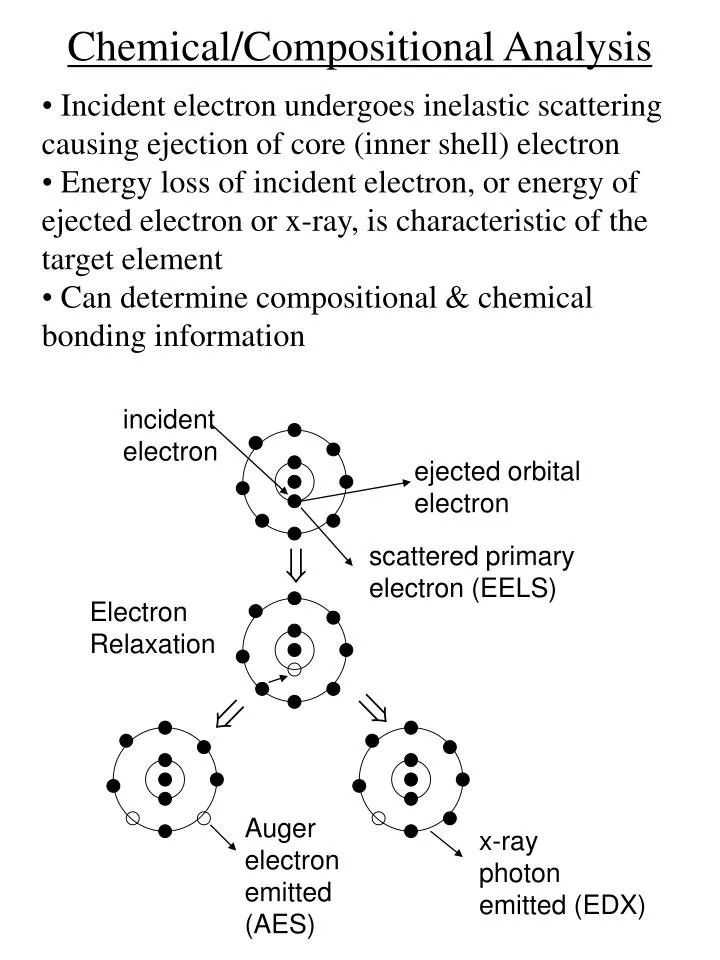
Chemical/Compositional Analysis. Incident electron undergoes inelastic scattering causing ejection of core (inner shell) electron Energy loss of incident electron, or energy of ejected electron or x-ray, is characteristic of the target element

E N D
Chemical/Compositional Analysis • Incident electron undergoes inelastic scattering causing ejection of core (inner shell) electron • Energy loss of incident electron, or energy of ejected electron or x-ray, is characteristic of the target element • Can determine compositional & chemical bonding information incident electron ejected orbital electron scattered primary electron (EELS) Electron Relaxation Auger electron emitted (AES) x-ray photon emitted (EDX)
Chemical/Compositional Analysis • Incident electrons undergo inelastic scattering with sample (X-rays, Auger electrons, secondary electrons) Electron Beam Secondary electrons (1-10 nm) Auger electrons (1 nm) Backscattered electrons (0.1 – 1 mm) X-rays (0.2 – 2 mm)
Electron Spectroscopy Electron, x-ray Spectroscopy EDX AES EELS PES Compositional Information Compositional & Chemical (Bonding) Information
EDX • EDX: Energy dispersive x-ray analysis • EDAX: Energy dispersive analysis of x-rays • EPM or EPMA: Electron probe micro-analysis • EDS: energy dispersive spectrometry • EMP: electron microprobe
EDX • Incident electron has sufficient energy (critical ionization energy) to eject a core shell electron From Ohring, Fig. 6-14(a) & (b), p. 278
EDX • Requires electron energy ~ keV Critical Ionization Energy for Pt
EDX • Outer shell electron fills the inner shell vacancy producing an x-ray photon (x-ray fluorescence) From Ohring, Fig. 6-14(c), p. 278
Kb La Ka EDX • X-ray nomenclature: • Ka : L → K • Kb : M → K • La : M →L Energy transition Terminating energy level • Letters denote principal quantum numbers (K: n = 1, L: n=2, etc.) adapted from Loretto, Fig. 2.3, p. 30
Kb La Ka EDX • Energy of emitted x-ray is determined by difference in electron energy levels : • hn = E(Ka) = EK- EL adapted from Loretto, Fig. 2.3, p. 30
EDX • X-ray energies are characteristic of the element From Ohring, Fig. 6-16, p. 281
EDX • Can identify the element from the x-ray lines emitted (Ti) From Ohring, Fig. 6-15(b), p. 280
EDX • X-ray detectors • EDS • Si p-i-n junction diode & MCA • Resolution ~ 150 eV • Based on energy of x-rays (EDS) From Williams, Fig. 1.16, p. 10
EDX • X-ray detectors • WDS • Uses Bragg reflection from crystal with known interplanar spacings to select l • Resolution ~ 5 eV • Based on l of x-rays from Schroder, Fig. 10.15, p. 669
EDX from Schroder, Table 10.1, p. 671 from Schroder, Fig. 10.16, p. 672
EDX • Can perform EDX using SEM or STEM (AEM) • Can produce elemental maps From Schroder, Fig. 10.4, p. 655
EDX • Can form compositional maps
EDX • EDX-SEM : • Lateral resolution ~ 1 mm • Depth resolution ~ 1 mm • EDX-AEM : • Lateral resolution ~ beam diameter (~0.5-2 nm) • Depth resolution ~ sample thickness (~2000 Å) Electron Beam Secondary electrons (1-10 nm) Auger electrons (1 nm) Backscattered electrons (0.1 – 1 mm) X-rays (0.2 – 2 mm)
EDX Quantification • Can determine amount of element present (to within ~ 0.1 at %)by measuring x-ray line intensity • Method 1: Calculation • Intensity of x-rays from a depth d is : • I = Ie(d)cswxe-md/cosq e dW/4p • Ie(d) = intensity of e-beam at depth d • c = atomic concentration • s = ionization cross-section • wx = x-ray yield (fluorescence yield) • = x-ray absorption coefficient q = detector angle wrt e-beam • e = detector efficiency • dW = detector solid angle
EDX Quantification • Method 2: Comparison with known standards • Compare x-ray intensity of sample with x-ray intensity from standard with known composition
EDX • X-ray emission competes with Auger process • Fluorescence yield is low below Na • EDX can detect elements above Na from Schroder, Fig. 10.14, p. 668
Can also use incident x-rays instead of electron beam • XRF: X-ray Fluorescence • XRFS: X-ray fluorescence spectroscopy
AES • Auger electron spectroscopy • Incident electron (few keV) ejects core electron from sample • Energy from electron transition is transferred to another electron (the Auger electron) causing it to be ejected incident electron ejected orbital electron scattered primary electron (EELS) Electron Relaxation Auger electron emitted (AES) x-ray photon emitted (EDX)
AES • Incident electron has sufficient energy to eject a core shell electron from Ohring, Fig. 6-14(a) & (b), p. 278
AES • Outer shell electron fills the inner shell vacancy causing ejection of Auger electron from Ohring, Fig. 6-14(d), p. 278
AES • Auger process requires 3 electrons (incident, core, Auger) • Can detect all elements except H & He from Ohring, Fig. 6-14(d), p. 278
AES • Auger nomenclature: • KLL Level of first ejected electron Initial level of Auger electron Level of electron that moves from outer to inner shell to fill electron vacancy
AES • Energy of Auger electron is determined by difference in electron energy levels : • E(KL1L2) = (EK- EL1) – (EL2+ f) Work function Energy released Energy required for Auger electron to escape surface
AES • Auger electron energies (~ 30 – 3000 eV) are characteristic of the element • Can detect all elements except H & He from Ohring, Fig. 6-17, p. 281
AES • Usually Auger signals, N(E), are differentiated, dN(E)/dE, to accentuate them from the background direct Auger spectra differentiated Auger spectra From Schroder, Fig. 10.10, p. 663
AES • Can identify the element from the AES spectrum Ti From Ohring, Fig. 6-15(c), p. 280
AES • Depth resolution determined by escape depth of electrons, < 20 Å • AES is a surface-sensitive technique; requires UHV • Depth profiling achieved using sputter gun from Yu & Cardona, Fig. 8.5, p. 420
AES • Lateral resolution ~ 10 - 50 nm (field emission source, scanning Auger) to 100 mm (non-scanning) • Cylindrical mirror analyzer (CMA) • Spectrometer resolution ~ 4 – 10 eV • Sensitivity ~ 0.1 – 1 at% • Quantification (10 % accuracy) achieved using calibrated standards from Ohring, Fig. 6-18, p. 284
EELS • Electron energy loss spectroscopy • Can examine energy loss of incident electron incident electron ejected orbital electron scattered primary electron (EELS) Electron Relaxation Auger electron emitted (AES) x-ray photon emitted (EDX)
EELS • Typical EELS Spectrum • Energy loss peak is characteristic of the elements DE = EK = binding energy of ejected electron Energy loss zero-loss peak (used as energy reference) energy loss due to inner shell ionization
EELS • EELS detector From Williams, Fig. 1.15, p. 9
EELS • EELS usually employed in TEM (AEM) From Williams, Fig. 1.2, p. 2
EELS • Fine structure is present in EELS spectrum • ELNES (energy loss near edge structure) • EXELS (extended energy loss spectroscopy) • Gives local atomic structure • chemical bonding information • atomic bond lengths (e.g., VCA) (EXELS) (ELNES)
EELS • EELS is complementary to EDX • More sensitive to low Z elements than EDX • Fine structure gives local atomic structure information
X-ray Absorption Spectroscopy • Can also observe fine structure in x-ray absorption • Measure transmission or fluorescence from sample as a function of incident x-ray photon energy • Core level excitations produce peaks in absorption or fluorescence • Fine structure in absorption edge gives chemical bonding information • XANES (x-ray absorption near-edge structure) or NEXAFS (near edge x-ray absorption fine structure • EXAFS (extended x-ray absorption fine structure) from C. Lamberti, Surf. Sci. Rep. 53 (2004) 1-197
X-ray Absorption Spectroscopy m(E)x = ln [Ii(E) / It(E) ] k = (2p/h) √ 2mo(hn – Eo) ; Eo = photoelectron binding energy
X-ray Absorption Spectroscopy • Synchrotron radiation is linearly polarized • Polarization-dependent EXAFS • gives information on bond orientation • can measure Da and Da in strained • layers
X-ray Absorption Spectroscopy • Can achieve surface sensitivity (~ 10 Å) by using grazing incidence geometry • Or detect Auger or photoelectrons (lower escape depth compared to fluorescence) • surface EXAFS (SEXAFS)
Photoemission Spectroscopy (PES) • X-ray photoelectron spectroscopy (XPS) • EDX and AES use incident electrons • XPS uses incident x-rays (few keV) to cause ionization (photoelectric effect) • Measure energy of ejected electron • = electron spectroscopy for chemical analysis (ESCA) • Ultraviolet photoelectron spectroscopy (UPS) • Uses uv photons From Schroder, Fig. 10.34, p. 702
PES • K.E. of ejected electron is characteristic of the element : • Ephotoelectron = hn- EB - incident photon energy binding energy of ejected electron (ionization energy) • Can detect elements above Li • Requires very good spectrometer for H, He
PES from Ibach and Luth, Fig. V.1, p. 125
PES • Typical XPS spectrum From Ohring, Fig. 6-15(d), p. 280
PES • XPS Advantages: • X-rays less prone to damage surfaces than electrons (e.g., electrons can reduce hydrocarbons on surface to carbon) • EB is sensitive to chemical surroundings (e.g., Si versus SiO2) • Typical applications are determination of electronic states (e.g., oxides, heterojunction band alignments)
ARXPS • Angle-Resolved XPS: • At grazing angles of detection only electrons from top surface region can escape • Surface-sensitive technique XPS detector path length of electrons is too large for escape
PEEM • PEEM: • Photoelectron emission microscopy • = photoelectron spectromicroscopy • Provides laterally resolved PES
Summary From Ohring, Fig. 6-15, p. 280