Download
1 / 63
650 likes | 771 Views
SPECIFIC CELLULAR RESPONSE. Paulina Roszkowska. The lymphoid system Main part - lymphocytes – differentiate from stem cells in the primary organs, migrate to secondary, recognize antigens by specific receptors. Central (primary) organs:

E N D
SPECIFIC CELLULAR RESPONSE Paulina Roszkowska
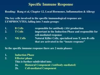
The lymphoid systemMain part - lymphocytes – differentiate from stem cells in the primary organs, migrate to secondary, recognize antigens by specific receptors • Central (primary) organs: * thymus - proliferation and maturation T lymphocytes, selection for tolerance to autoantigens * bone marrow –proliferation and maturation B lymphocytes • Secondary (peripheral):an environment in which mature lymphocytes T i B interact with antigen, cooperate with phagocytes and accessory cells * spleen – responds to blood-borne antigens * lymph nodes – lymph-borne antigens absorbed via skin, * mucosal tissue - MALT (GALT, BALT), .. – protects the mucosal surface • there is continuous lymfocyte traffic - from the blood stream into lymphoid tissues and back again
Non-specific and Specific Immunity: Contrasts Non-specific (natural, native, innate) system in place prior to exposure to antigen can be enhanced after exposure to antigen through effects of cytokines lacks discrimination among antigens Specific (acquired, adaptive) is induced by specific antigen Is activated later than non-specific has memory Learnt by experience Enhanced by second exposure Is poorly effective without innate immunity The non-specific and specific immune systems interact with each other!
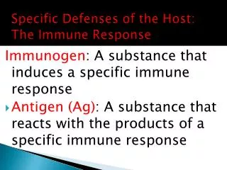
Antigen (Ag) A substance that can induce a specific immune response and react with the products of it. • Epitope – single determinant of antigen, binds with paratope of antibody
Feature of antigens • Immunogenicity – ability of recognition of the antigen and production of cellular or humoral response • Antigenicity - ability of raction with products of cellular or humoral response • Specificity – reaction of antibody or specific lymphocytes with epitope
Hapten A substance that is non-immunogenic but which can react with the products of a specific immune response. Haptens are small molecules which could never induce an immune response by themselves but they can when coupled to a carrier molecule. Haptens have the property of antigenicity but not immunogenicity.
Superantigens • proteins produced by pathogens, which are not processed by antigen presenting cells but can binds to variable region of β chain on TCR of T cells and to MHC class II on antigen presenting cells (APC) and activate Tcell • Large numbers of activated T cells release cytokines having pathological effects
Antigenic peptides must normally be processed in order to triger the TCR recognition in MHC groove. It gives: low frequency of antigen- antigen specific cells Superantigens (e.g.staphyloccocal toxins) are not processed but bind directly to MHC II only. It gives: high frequency of antigen- antigen specific cells: polyclonal response
ANTIGEN + LYMPHOCYT = IMMUNE RESPONSE but • different antigens • react with different cells of immune system • and cause different response
Leucocytes Adaptive and innate immunity depends upon LEUCOCYTES Innate immunity is mediated largely by GRANULOCYTES Adaptive immunity mediated by LYMPHOCYTES The growth, development and activities of granulocytes and lymphocytes are interconnected and co-operative.
CLP T CELLS B CELLS Common lymphoid precursor T B Th CTL PC Activate B cells and macrophages T HELPER CELLS Kill virus- infected cells CYTOTOXIC T LYMPHOCYTES Produce antibodies PLASMA CELLS Lymphocyte subsets
Lymphocytes 20-40% of whole leucocytes in blood, morphologically different: • T - 70% -TCR receptor + CD 3 • Th 1, Th 2 – helper • 50-55% • CD4 – co-receptor for MHC-II, 95% small lymphocytes • Tc – cytotoxic • 20-25% • CD8 - MHC-I – 50% small lymphocytes • Ts – suppressor ? (CD8 but also CD4) • B - 10-20% -CD19 CD20 CD21 + -IgM, IgD, MHC-II receptors, only few have IgG,IgA,IgE expression • NK - (CD56, CD57) - large granular
Lymphocytes • express characteristic surface markers detected by monoclonal antibodies- cluster of differentiation antigens CD (> 150 is known now) • Markers are responsible for phenotype and biological function: • T – CD2- binds sheep blood cells • CD3- signal tranduction • release cytokines: • Th1: Il-2, IFN , TNF • Th2: Il-2, Il-4, Il-5, Il-6, Il-9 Il-10, Il-13 • possess receptors for: • cytokins, • MHC, • Fc (CD16, CD64), • C, • integryns (CD2 - LFA), selektyns (CD62 - E, L, P) • T or B actiovation releases additional expression of surface markers, antigens
Lymphocyte antigen receptors T and B cells are essentially inactive until they encounter antigen. T and B cells express ANTIGEN RECEPTORS Lyc The B cell antigen receptor is a membrane-bound antibody SURFACE IMMUNOGLOBULIN B The T cell antigen receptor IS NOT membrane bound antibody but a distinct molecule T CELL ANTIGEN RECEPTOR T Each antigen receptor binds to a different antigen
What Does The B Cell Immunoglobulin (Ig) Receptor Recognize? • Proteins (conformational determinants, denatured or proteolyzed determinants) • Nucleic acids • Polysaccharides • Some lipids • Small chemicals (haptens)
Uniqueness of B Cells • Express both immunoglobulin (Ig) and class II MHC on cell surface • Capable of producing antibody of same specificity as that of its surface Ig AND • Capable of functioning as an antigen presenting cell
What Does the T Cell Receptor (TCR) Recognize? Only fragments of proteins (peptides) associated with MHC molecules on surface of cells • Helper T cells (Th) recognize peptide associated with MHC class II molecules • Cytotoxic T cells (Tc) recognize peptide associated with MHC class I molecules • Every TCR on an individual T cell has one specificity
T cell receptor (TCR) • The specificity for immune responses resides in the T cell receptor (TCR) • recognizes pathogen (antigen)-derived peptides bound to major histocompatibility complex (MHC) molecules expressed on the surface of nucleated cells –APC (antigen presenting cells).
Types of antigens: • T –dependent antigen • need recognition by both T and B lymphocytes to produce antibodies, • most antigens, • mainly proteins, • humoral and cellular response • T-independent antigen • recognition by B lymphocytes B, • mainly polysaccharides: capsule of S.pneumoniae, H.influenzae, N.miningitidis • only antibodies are produced, no cellular response
Location of Pathogen Determines Which T Cell Population Responds • Cytosolic: • cells harboring pathogens in the cytosol are recognized and killed by cytotoxic T cells (Tc) that express CD8 • Vesicular: • cells are recognized by a subpopulation of helper T cells (Th1) that express CD4, which enable the infected cell to kill the pathogen • Extracellular: subpopulation of helper T cells (Th2) that express CD4
Location of Pathogen Determines Which T Cell Population Responds Extracellular - site of most bacteria as: -T-independent antigen -T-dependent antigen subpopulation of helper T cells (Th2) that express CD4 elicits antibody (humoral) response Intracellular reside in two intracellular compartments: Cytosolic: viruses and some bacteria cytosol and nucleus connected via nuclear pores cells harboring pathogens in the cytosol are recognized and killed by cytotoxic T cells (Tc) that express CD8 Vesicular: some bacteria, some parasites membrane-bound entities (endoplasmic reticulum, endosomes, lysosomes, Golgi apparatus) cells are recognized by a subpopulation of helper T cells (Th1) that express CD4, which enable the infected cell to kill the pathogen - elicits cell-mediated response
T Y Cell surface peptides of Ag presented by cells that express MHC antigens Soluble native Ag Soluble peptides of Ag Cell surface peptides of Ag Cell surface native Ag ExogenousAntigens must be processed in order to be recognised by T cells ANTIGEN PROCESSING T cell response No T cell response No T cell response No T cell response No T cell response
Leukocyte Migration and Localization • Bone marrow and thymus (primary lymphoid tissues) • produce B cells and T cells • B cells and T cells recirculate through spleen and lymph nodes (secondary lymphoid tissues)
Lymphocyte Recirculation • Antigen presenting cells (APC) pick up antigen and migrate to secondary lymphoid tissues and interact with T cells and B cells • Secondary lymphoid tissues (lymph nodes, spleen) main sites where lymphocytes encounter antigen • Frequency of lymphocytes having a receptor specific for a given antigen is low
T cell T cell T cell B cell B cell B cell APC B cell T cell Leukocyte Migration and Localization Bone marrow Macrophage Thymus Dendritic cell Naive lymphocytes Spleen and lymph nodes Tissues Primed lymphocytes
APC – Antigen Presenting Cells • Heterogenic population: • Langerhans cells – present to Th1 • B cells– present to Th2 • macrophages– phagocytes • Role of APC: • antigen processing (degradation into small peptides), • binding with MHC-II and • presentation to T CD4 • All cells can present antigen to T CD8 - posses MHC-I
SELF MHC RESTRICTION • In order for a T cell to recognize and respond to a foreign protein antigen, it must recognize the MHC on the presenting cell as self MHC. • Helper T cells recognize antigen in context of class II self MHC. • Cytolytic T cells recognize antigen in context of class I self MHC. • The process whereby T cells become restricted to recognizing self MHC molecules occurs in the thymus.
Class I MHC Molecules expressed on surface of all nucleated cells recognized by TCR of cytotoxic T cells CD8 binds to class I MHC-peptide complex source of peptide is cytosolic compartment Class II MHC Molecules expressed on surface of some nucleated cells, mainly antigen presenting cells (APC) recognized by TCR of helper T cells CD4 binds to class II MHC-peptide complex source of peptide is vesicular compartment
Different cellular pathways for association of peptide with MHC class I and class II molecules
Viral protein is made oncytoplasmic ribosomes Plasma membrane Class I MHC Pathway Peptide is presented by MHC-I to CD8 cytotoxic T cell Globular viral protein - intact Peptide passes with MHC from Golgi body to surface Proteasome degrades protein to peptides rER Peptide associates with MHC-I complex Peptide transporter protein moves peptide into ER MHC class I alpha and beta proteins are made on the rER Peptide with MHC goes to Golgi body Golgi body
Peptide MHC-II complex is presented to CD4 helper T cell Globular protein CD4 helper T cell Endosome Endosome fuses with plasma membrane Endocytosis Fusion of endosome and exocytic vesicle Immunodominant peptide binds to class II MHC Lysosome Exocytic vesicle fuses with endosome releasing Ii from αβ dimer Protein is processed to peptides in endosome or lysosome Golgi body Class II MHC Synthesis 3 chains: α,β and Ii α β Ii Endoplasmic reticulum Class II MHC Pathway
Immune response Different cells cause different response
T cells + antigen memory T cells memory effector T cells Tc lymphocytes Th macrophages NK lymphocytes CELLULAR RESPONSE B cells + antigen humoral antigen T-independent antigen memory B cells memory effector B cells -plasma cells antybodies HUMORAL RESPONSE Immunologic responce
Main phases of the immune response: • induction phase • recognition of antigen • central phase • activation, • clonal selection and prolipheration of T and B lymphocytes • effector phase • elimination of antigen mediated by antibodies and effector cells
Cell medited specific responseImmune response phases: • INDUCTION • recognition of antigen by T lymphocyte, • central phase • the main function of CD4 cell- regulation by cytokines • EFFECTOR • elimination of antigen, pathogen: • macrophages activation (Th1 - IFN)) – better destroing of intracellular bacteria: mycobacteria, brucella, listeria • cytotoxicity – T CD8 (Th - Il-2 + APC - Il-12) cell-cell interaction against whole cells: virus-infected, tumour, transplant cells..by perforins, Fas ligand • ADCC- antibody dependent cell citytoxity –K cells, receptor Fc (CD16), bind antigens conected with cell • LAK – Il-2 activated lymphocytes NK ? TcCD8, NK activated,
viruses in cytosol, MHC class I pathway, Tc response • extracellular bacteria, MHC class II pathway, Th2 response, Ab formation • intracellular bacteria, MHC class II pathway, Th1 response
Naive Th cells Short-term stimulation Long term Memory cells Chronic stimulation IFNγ IL-2 IL-12 ThM cell Th2 cell Th1 cell ThO cell ThP cell IL-4 IFNγ IL-2 IL-4 IL-5 IL-10 IL-2 IL-2 IL-4 IL-5 IL-6 IL-10 Naïve Th Cells Can Differentiate Into Th1 or Th2 Cells
- IFN- - IL-10 Th1 Th2 Balanced response - - IL-10 Th1 IFN- Th2 Th2 Th1 Th2 Dominant Th1 Dominant Counter regulation of Th cell subsets
Inhibits proliferation IL-10 Inhibits production Th1 cell Th2 cell IL-4 IL-5 IFNγ Activates Activates B cell Eosinophil Mast cell Macrophage Antibodies (including IgE) Functions of Th1 and Th2 Cells
Th1 -Th cells are a subset of T cells that express a unique antigen on their surface called CD4. -A subpopulation of Th1 cells, is the primary defense against intracellular pathogens (inside vesicles). • Th1 cells recognize antigen from the pathogen that are expressed on the surface of infected cells • release cytokines that activate the infected cell. • Once activated, the infected cell can then kill the pathogen. • For example, Mycobacterium tuberculosis, infects macrophages but is not killed because it blocks the fusion of lysosomes with the endosomes. Th1 cells that recognize M. tuberculosis antigens on the surface of an infected macrophage can secrete cytokines that activate macrophages.
lysosome Macrophage Th1 cell Macrophage antigen mycobacteria Activated infected macrophage Infected macrophage Helper (Th1) T Cells
Th cell Invading agent Antigen presentation Activated macrophage Activate Macrophage Macrophage Macrophage Cytokines Anti-microbial functions Cytokines Lymphokines Anti-tumor functions Central Role of Macrophages in Natural and Specific Immunity • Involved in initial defense and antigen presentation and have effector functions
Cytotoxic T lymphocytes (CTL) • CTLs are a subset of T lymphocytes that express a unique antigen on their surface called CD8. • These cells recognize antigens from the pathogen that are displayed on the surface of the infected cell and kill the cell. • CTLs kill by cytotoity and inducing apoptosis in the infected cell
A B Cell expresses viral antigens Cytotoxic T cell C Virus infects cell Infected cell is killed by cytotoxic T cell by activation of nucleases that cleave host and viral DNA Cytotoxic (Tc) T Cells