Download
1 / 48
510 likes | 1.26k Views
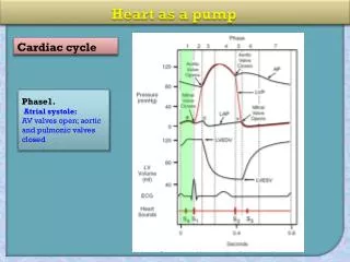
Heart as a Pump . Outline . Overview of heart anatomy and function Cardiac cycle . Volume-‐Pressure Diagram . Cardiac Output and Venous Return Regulation of Cardiac Output . Learning Objectives . Describe the cardiac cycle by explaining Fig. 9-6 .

E N D
Outline Overview of heart anatomy and function Cardiac cycle Volume-‐Pressure Diagram Cardiac Output and Venous Return Regulation of Cardiac Output
Learning Objectives • Describe the cardiac cycle by explaining Fig. 9-6 in Guyton and Hall • Analyze ventricular pumping with a volume ‐ pressure diagram • Understand cardiac output and venous return - quantitatively know cardiac output • Know how cardiac output is regulated - Frank- Starling mechanism and autonomic regulation
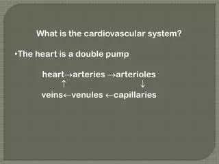
Cardiovascular System Note, right side is the right side of the person or animal. Two pumps in the heart: Right side pumps blood through the lungs Left side through the peripheral organs Each side has an atrium and a ventricle Atrium is a primer pump for the ventricle Ventricle supplies the main pumping force
Heart Anatomy Atrioventricular valves: tricuspid (right) and mitral (left) Semilunar valves: pulmonary (right) and aortic (left)
Cardiac Cycle • The cardiac cycle includes the events that occur from the beginning of one heartbeat to the beginning of the next • The cardiac cycle consists of two periods: - Diastole - period of relaxation when the heart fills with blood - Systole - period of contraction
Beginning just after a ventricular contraction Semilunar valves closed AV valves opened Diastole: Passive ventricular filling. The AV valves open and blood flows into the relaxed ventricles, accounting for most of the ventricular filling.
Semilunar valves closed AV valves opened Diastole: Active ventricular filling. Semilunar valves closed AV valves opened Diastole: Passive ventricular filling.
Semilunar valves closed AV valves opened Diastole: Active ventricular filling. The atria contract and complete ventricular filling.
Semilunar valves closed AV valves closed Systole: Period of isovolumic contraction. Semilunar valves closed AV valves opened Diastole: Active ventricular filling.
Semilunar valves closed AV valves closed Systole: Period of isovolumic contraction. Ventricular contraction causes the AV valves to close, which is the beginning of ventricular systole. The semilunar valves were closed in the previous diastole and remain closed during this period.
Semilunar Semilunar valves opened valves closed AV valves closed AV valves closed Systole: Period of Systole: Period of ejection. isovolumic contraction.
Semilunar valves opened AV valves closed Systole: Period of ejection. Continued ventricular contraction pushes blood out of the ventricles, causing the semilunar valves to open.
Semilunar valves opened AV valves closed Systole: Period of ejection. Semilunar valves closed AV valves closed Diastole: Period of isovolumic relaxation.
Semilunar valves closed AV valves closed Diastole: Period of isovolumic relaxation. Blood flowing back toward the relaxed ventricles causes the semilunar valves to close, which is the beginning of ventricular diastole. Note that the AV valves closed, also.
Semilunar valves closed AV valves closed Diastole: Period of Semilunar valves closed isovolumic relaxation. AV valves opened Diastole: Passive ventricular filling.
Mechanical Events: The Cardiac Cycle
The Cardiac Cycle • Cardiac cycle refers to all events associated with blood flow through the heart from the start of one heartbeat to the beginning of the next • During a cardiac cycle • Each heart chamber goes through systole and diastole • Correct pressure relationships are dependent on careful timing of contractions
Phases of the Cardiac Cycle • Atrial diastole and systole - • Blood flows into and passively out of atria (80% of total) • AV valves open • Atrial systole pumps only about 20% of blood into ventricles • Ventricular filling: mid-to-late diastole • Heart blood pressure is low as blood enters atria and flows into ventricles • 80% of blood enters ventricles passively • AV valves are open, then atrial systole occurs • Atrial systole pumps remaining 20% of blood into ventricles
Phases of the Cardiac Cycle • Ventricular systole • Atria relax • Rising ventricular pressure results in closing of AV valves (1st heart sound – “lubb”) • Isovolumetric contraction phase • Ventricles are contracting but no blood is leaving • Ventricular pressure not great enough to open semilunar valves • Ventricular ejection phase opens semilunar valves • Ventricular pressure now greater than pressure in arteries (aorta and pulmonary trunk)
Phases of the Cardiac Cycle • Ventricular diastole • Ventricles relax • Backflow of blood in aorta and pulmonary trunk closes semilunar valves (2nd hear sound - “dubb”) • Dicrotic notch – brief rise in aortic pressure caused by backflow of blood rebounding off semilunar valves • Blood once again flowing into relaxed atria and passively into ventricles
Normal Volume of Blood in Ventricles • After atrial contraction, 110-120 ml in each ventricle (end-diastolic volume) • Contraction ejects ~70 ml (stroke volume output) • Thus, 40-50 ml remain in each ventricle (End‐ systolic volume) • The fraction ejected is then ~60% (ejection fraction)
Left Ventricle Volume-‐Pressure Curve Be able to use these pressure and volume values Aortic valve closes Aortic valve opens Mitral valve opens Mitral valve closes End-systolic volume End-diastolic volume
Preload and Afterload • Preload - tension on muscle when it begins to contract (end-diastolic pressure) • Afterload - load against which the muscle exerts its contractile force, which is the pressure in the artery leading from the ventricle. Phase III on volume-pressure diagram
Cardiac Output and Venous Return • Cardiac output is the quantity of blood pumped into the aorta each minute. Cardiac output = stroke volume x heart rate • Venous return is the quantity of blood flowing from the veins to the right atrium. • Except for temporary moments, the cardiac output should equal the venous return
Normal Cardiac Output • Normal resting cardiac output: - Stroke volume of 70 ml - Heart rate of 72 beats/minute - Cardiac output ~ 5 litres/minute • During exercise, cardiac output may increase to > 20 liters/minutes • You should be able to get stroke volume and heart rate from volume-‐pressure curves and ECG recordings, respectively
Cardiac Output • Stroke Volume = the vol of blood pumped by either the right or left ventricle during 1 ventricular contraction. SV = EDV – ESV 70 = 125 – 55 CO = SV x HR 5,250 = 70 ml/beat x 75 beats/min CO = 5.25 L/min
Cardiac Output • Regulation of Stroke volume • Preload: Degree of stretch of heart muscle (Frank-Starling) – greatest factor influencing stretch is venous return (see Below) • Contractility – Strength of contraction Increased Ca2+ is the result of sympathetic nervous system
Cardiac Output • Other chemicals can affect contractility: - Positive inotropic agents: glucagon, epinephrine, thyroxine, digitalis. - Negative inotropic agents: acidoses, rising K+, Ca2+ channel blockers. Afterload: Back pressure exerted by arterial blood. Regulation of Heart Rate • Autonomic nervous system • Chemical Regulation: Hormones (e.g., epinephrine, thyroxine) and ions.
Regulation of Cardiac Output • Frank-Starling Mechanism -‐ Cardiac output changes in response to changes in venous return. • Autonomic control -‐ Control of heart rate and strength of heart pumping by the autonomic nervous system.
Chemical Regulation of the Heart • The hormones epinephrine and thyroxine increase heart rate • Intra- and extracellular ion concentrations must be maintained for normal heart function
Regulation of Stroke Volume • SV: volume of blood pumped by a ventricle per beat SV= end diastolic volume (EDV) minus end systolic volume (ESV); SV = EDV - ESV • EDV = end diastolic volume • amount of blood in a ventricle at end of diastole • ESV = end systolic volume • amount of blood remaining in a ventricle after contraction • Ejection Fraction - % of EDV that is pumped by the ventricle; important clinical parameter • Ejection fraction should be about 55-60% or higher
Factors Affecting Stroke Volume • EDV - affected by • Venous return - vol. of blood returning to heart • Preload – amount ventricles are stretched by blood (=EDV) • ESV - affected by • Contractility – myocardial contractile force due to factors other than EDV • Afterload – back pressure exerted by blood in the large arteries leaving the heart
Frank-Starling Law of the Heart • Preload, or degree of stretch, of cardiac muscle cells before they contract is the critical factor controlling stroke volume; EDV leads to stretch of myocardium. • preload stretch of muscle force of contraction SV • Unlike skeletal fibers, cardiac fibers contract MORE FORCEFULLY when stretched thus ejecting MORE BLOOD (SV) • If SV is increased, then ESV is decreased!! • Slow heartbeat and exercise increase venous return (VR) to the heart, increasing SV. • VR changes in response to blood volume, skeletal muscle activity, alterations in cardiac output • VR EDV and in VR in EDV • Any in EDV in SV • Blood loss and extremely rapid heartbeat decrease SV.
Frank-Starling Mechanism The force of cardiac muscle contraction increases as the muscle stretches, within limits. Due to more optimal overlap of actin and myosin filaments during stretch - same in skeletal muscle So, with increase venous return and increased stretching, the force of contraction increases and the stroke volume increases. Moreover, stretching of the SA node increasing the firing rate of the pacemaker(increasing heart rate).
Frank-‐Starling Summary: within physiological limits, the heart pumps all the blood that returns to it from the veins. Venous return increases when there is an increase in the blood flow through peripheral organs. So, peripheral blood flow is a major determinant of cardiac output
Extrinsic Factors Influencing Stroke Volume • Contractility is the increase in contractile strength, independent of stretch and EDV • Referred to as extrinsic since the influencing factor is from some external source • Increase in contractility comes from: • Increased sympathetic stimuli • Certain hormones • Ca2+ and some drugs • Agents/factors that decrease contractility include: • Acidosis • Increased extracellular K+ • Calcium channel blockers
Effects of Autonomic Activity on Contractility • Sympathetic stimulation • Release norepinephrine from symp. postganglionic fiber • Also, EP and NE from adrenal medulla • Have positive ionotropic effect • Ventricles contract more forcefully, increasing SV, increasing ejection fraction and decreasing ESV • Parasympathetic stimulation via Vagus Nerve -CNX • Releases ACh • Has a negative inotropic effect • Hyperpolarization and inhibition • Force of contractions is reduced, ejection fraction decreased
Contractility and Norepinephrine • Sympathetic stimulation releases norepinephrine and initiates a cyclic AMP 2nd-messenger system Figure 18.22
Preload and Afterload Figure 18.21
Effects of Hormones on Contractility • Epi, NE, and Thyroxine all have positive ionotropic effects and thus contractility • Digitalis elevates intracellular Ca++ concentrations by interfering with its removal from sarcoplasm of cardiac cells • Beta-blockers (propanolol, timolol) block beta-receptors and prevent sympathetic stimulation of heart (neg. chronotropic effect)
Autonomic Control of Cardiac Output Sympathetic increases cardiac output Can increase heart rate 70 to 180-200 BPM Can double force of contraction Sympathetic nerves release norepinephrine Believed to increase permeability of Ca2+ and Na+. Parasympathetic (vagal) decreases cardiac output Can decrease heart rate to 20-40 BPM Can decrease force of contraction by 20-30% Parasympathetic nerves release acetylcholine Increases permeability to K+
Cardiac Output and Peripheral Resistance Increasing the peripheral resistance decreases cardiac output. arterial pressure total peripheral resistance cardiac output =
Other Factors Affecting Cardiac Output • Age • Gender • Exercise/body temperature