Download
1 / 20
220 likes | 1.39k Views
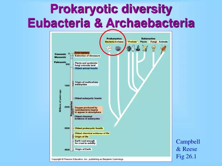
Prokaryotic diversity Eubacteria & Archaebacteria. Campbell & Reese Fig 26.1. Bacteria. one-celled to simple colonies cells prokaryotic metabolism diverse. Mycobacterium paratuberculosis. Archaeabacteria - extremophiles. one-celled to simple colonies cells prokaryotic

E N D
Prokaryotic diversityEubacteria & Archaebacteria Campbell & Reese Fig 26.1
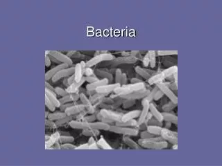
Bacteria • one-celled to simple colonies • cells prokaryotic • metabolism diverse Mycobacterium paratuberculosis
Archaeabacteria - extremophiles • one-celled to simple colonies • cells prokaryotic • chemistry different than Bacteria • metabolism diverse, “primitive” Grand prismatic pool, Yellowstone NP
Prokaryotes vs. Eukaryotes cells small (1-5 μm diameter) unicellular no nucleus or organelles cell wall cells large (10-100 mm) unicellular or multicellular nucleus and organelles different cell wall when present C&R Fig 27.2
Prokaryote shape rod-shaped (bacilli) spherical (cocci) helical
Very small • cells usually small (1-5 μm diameter) • largest are Cyanobacteria and, • Thiomargarita namibiensis, "Sulfur Pearl of Namibia" T. namibiensis next to a fruit fly 1 mm
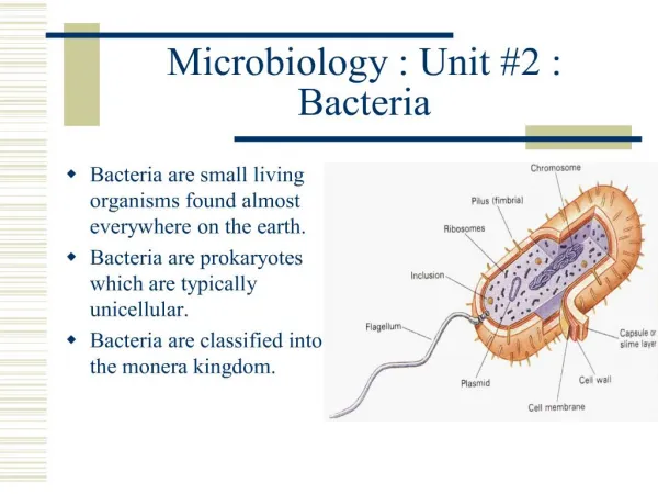
Prokaryotic cell walls maintain shape, protection complex chemically – peptidoglycan and lipids many antibiotics target this special chemistry Fig 27.5
Internal cellular structure one double stranded chromosome of DNA in the form of a ring smaller rings of DNA called plasmids specialized membranes for metabolic purposes no membrane-bound organelles aerobicprokaryote photosynthetic prokaryote
Movement Flagella (bacterial) Chemical gliding Link to Animated bacterial movement towards attractant Bacterial tumble movie
Asexual reproduction Binary fission Growth of Pneumococcus Time lapse over 2 hours Resistant spores
Sexual change No real sexual reproduction transformation - the uptake of genes from the surrounding environment transduction - transfer of genes from viruses to prokaryotes conjugation - direct transfer of genes from prokaryote to prokaryote
Nitrogen fixation Convert atmospheric nitrogen into biological form used in proteins and nucleic acids Anabaena, a photoautotroph, can also fix nitrogen. Heterocysts – cells specialized to carry out the process
Metabolism and oxygen obligate aerobes - oxygen required facultative aerobes - use oxygen when available but not required obligate anaerobes - poisoned by oxygen
Archaebacteria most research has focused on their ecology rather than phylogeny extreme halophiles extreme thermophiles methanogens
Ecolgical impacts of prokaryotes decomposers - recycle nutrients from dead organisms pathogens - cause human disease mutualists - live closely with another organism and both benefit
Economic roles of prokaryotes pathogens - cause human disease fermentation – vinegar, yogurt, cheese genetically engineered - insulin and interferon bioremediation – remove environmental contaminants source of unique compounds - T. aquaticus DNA polymerase
Are viruses alive? Virus structure nucleic acids “genes” (DNA or RNA) protein covering Other virus pictures (Electron micrographs)
Virus reproduction Require metabolic capabilities of a host cell Specific to host species/tissue/cell Recognize host cell surface Genes enter host Host follows instructions to build new virus Virus escapes
Virus diversity Many viruses In some ways more closely related to hosts than to each other