Download
1 / 54
540 likes | 712 Views
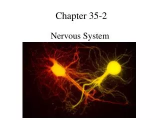
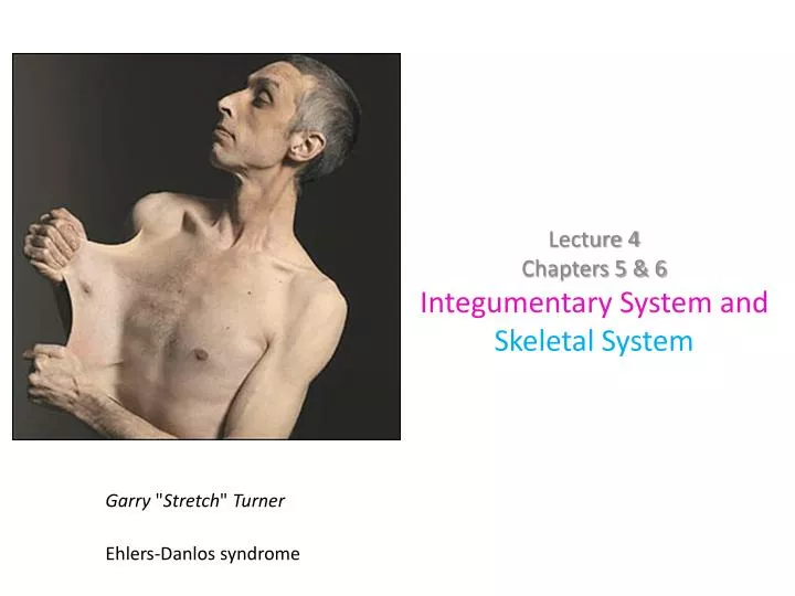
Lecture 4 Chapters 5 & 6 Integumentary System and Skeletal System. Garry " Stretch " Turner. Ehlers- Danlos syndrome. Body Tissues. Tissue membranes : consist of: Thin sheets of epithelium and c.t . that: 1. cover body surfaces or 2. line body cavities. Tissue Membranes.

E N D
Lecture 4 Chapters 5 & 6 Integumentary System and Skeletal System Garry "Stretch" Turner Ehlers-Danlos syndrome
Body Tissues • Tissue membranes: consist of: Thin sheets of epithelium and c.t. that: 1. cover body surfaces or 2. line body cavities.
Tissue Membranes • Tissue membranes - four major types: 1) serous membrane – lines a body cavity that does not open to the outside. e.g., thoracic (chest) cavity abdominopelvic cavity
Tissue Membranes • 2) mucous membrane – lines a body cavity or tube that opens to the outside. e.g., mouth & nasal cavities digestive, respiratory, urinary and reproductive tracts Composed of epithelial layer with embedded mucus producing cells (goblet cells) overlying a layer of loose c.t.
Tissue Membranes • 3) synovial membrane – lines a joint cavity. • Produces synovial fluid that cushions/lubricates • 4) cutaneous membrane – external body covering (i.e.skin)
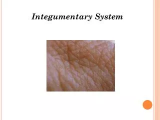
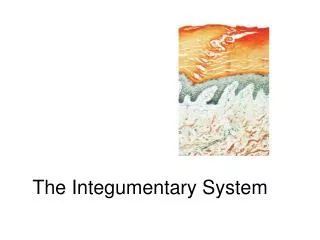
Integumentary System (skin) Organ system=2 or more tissues working together to perform some function A. Functions of the integument 1. Protection 2. Prevent H2O loss (keratin[protein], sebum [oil]) 3. Excretion (waste in sweat) 4. Temperature regulation (evaporation of sweat, vasodilatation and constriction) 5. Synthesis of vitamin D in sunlight 6. Sensation (touch, pressure, pain, temperature)
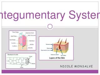
Skin Structure: Composed of 2 general layers: epidermis and dermis B. Layers 1. Epidermis– stratified squamous epithelium, keratinized (protein keratin, waterproof) epithelium. a. Has 4 to 5 layers, the superficial layer, stratum corneum is dead! b. Stratum basale (germinativum) is the bottomlayer that grows (mitosis) and contains melanocytes.
Superficial layer Basal layer Stratum=layer
Structure of the skin (cont) 2. Dermis – irregular, dense connective tissue 3. Subcutaneous (hypodermis)– underlying adipose and loose connective tissue, not really part of the true skin.
C. Specialized structures (accessory organs of skin) 1. Hair – derived from epidermis a. Grows from the hair follicle b. Serves as insulation in animals, the arrectorpilli muscles make goose bumps.
Accessory Organs of the skin (Cont) 2. Glands – derived from epidermis. a. Sebaceous (oil sebum) usually with the hair follicle. They lubricate the hair shaft, and waterproof the skin. Keeps skin soft
b. Sudoriferous (sweat) glands 1. Eccrine (Merocrine)-cools the body via evaporation, excretes water salt and waste (urea) at the surface 2.Apocrine- in axillae (armpit) and pubic regions develop at puberty and release secretions when excited/ anxious release pheromones.
Accessory organs of the skin (cont.) c. Sensory receptors – derived from structures in the dermis. 1. meisnner’scorpuscle for touch, 2. pacinian corpuscle for pressure Temperature and pain receptors are also present).
Accessory organs of the skin (cont) d. Nails – derived from the epidermis. -Rich in keratin =hard structure
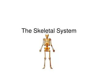
Skeletal System Andreas Vesalius
VII. Skeletal system • A. Functions of the Skeleton – • Support-site of attachment of soft tissues • protection - delicate tissues surrounded by bone • levers for movement- transmit forces from muscles • blood formation – produced in red marrow • calcium and phosphate storage
B. Classification of Bone (by type) Compact–dense, on bone surface, shaft and head of long bones. Spongy (cancellous) – lines marrow cavity in long bones, heads, middle part of short, flat, irregular bones, filled with redmarrow.
Classification of bone (by shape) 1. Long bones – appendages, all bones of limbs except wrist (carpals) and ankles (tarsals). 2. Short bones – wrist (carpals) and ankle (tarsals). 3. Flat bones– cranium (top of skull), sternum, ribs. 4. Irregular bones – vertebrae, pelvis, facial bones 5. Sesamoid-form inside tendon (knee cap) shaped like a sesame seed 6. Sutural (wormian)- islands of bone form between sutures in skull
C. Anatomy of a Long Bone 4. Spongy Bone = houses Red bone marrow 1. Epiphysis = expanded area at ends of long bone 5. Epiphysial line or plate =thin line of cartilage (line) or bone (plate) dite of bone growth lengthwise 3. Compact bone = outer layer of bone 6. Medullary cavity – hollow cavity, holds yellow bone marrow in life. 2. Diaphysis =long tubular shaft 7. Periosteum = fibrous cover attaches tendons and ligaments 8. Articular cartilage
D. Bone Growth and Remodeling 1. Intramembranous bone development – skull and clavicle. a. Fibrous connective tissue present only 2 months after conception is the template that is (ossified) converted to bone. This is not complete at birth and the “soft spots” fontanels remain for a time (up to 1 ½ years) after birth.
Cells of Bone • Osteoblasts: produces (deposits) new bone and assist in repair of bone • Osteoclast: breaks down old bone and assist in repair of bone • Osteocytes: mature bone cells help maintain bone
Bone Formation (cont) b. Intracartilaginous – within hyaline cartilage 1.) All the skeleton below the base of the skull except the clavicles. 2.) “Bones” first are formed of hyaline cartilage by about 3 months of age and begin ossification or the conversion to bone 3.) A Center of primary ossificationin long bones develops in the center of the diaphysis (shaft). 4.) Secondary centers of ossification develop later in each of the two epiphyses (heads). 5.) The bone head and shaft are separated at each end by a cartilage growth plate called the epiphyseal platewhile the centers convert hyaline cartilage to bone.
6.) Ossification in the primary center spreads toward the end of the shaft while ossification in the secondary centers spreads toward the shaft. 7.) A long bone has the epiphyseal plate of cartilage can continue to grow in length . That is about 18 years in females and 21 in males. 8.) When cartilage growth is no longer faster than bone formation in the primary and secondary centers, the epiphyseal plate is converted to bone leaving the epiphyseal line. This epiphyseal closure means the bone can no longer grow in length. It is still capable of growing in diameter. (appositional growth)
Appositional growth is from osteoprogenitor cells under the periosteum
E. Organization of the Skeleton 1. Axial – head, spine, chest. a. Cranium 1.) Frontal (1). 2.) Parietal (2). 3.) Temporal (2). 4.) Occipital (1). 5.) Sphenoid (1). 6.) Ethmoid (1). b. Face 1.) Zygomatic (2). 2.) Lacrimal (2). 3.) Nasal (2). 4.) Vomer (1). 5.) Inferior nasal conchae (2).
6.) Maxillae (2). 7.) Palatine (2). 8.) Mandible (1). c. Hyoid d. Trunk – 1.) vertebral column. a.) Cervical (7). b.) Thoracic (12). c.) Lumbar (5). d.) Sacrum (1) composed of 5 fused vertebrae. e.) Coccyx (1) composed of 4 fused vertebrae. 2.) Sternum (1) composed of manubrium, body, and xiphoid process.
3.) Ribs – 24, (12 pairs). a.) True ribs, first 7 pairs. b.) False ribs, last 5 pairs, last 2 pairs of false ribs are floating. 2. Appendicular Skeleton – pectoral girdle, arms pelvic girdle, legs.
a. Upper extremities 1.) Pectoral girdle a.) Scapulae (2). b.) Clavicles (2). 2.) Arm (#/arm) a.) Humerus (1). b.) Radius (1). c.) Ulna (1). d.) Carpals (8).
e.) Metacarpals (5). f.) Phalanges (14), 2 in thumb, 3 in each digit.
b. Lower Extremity 1.) Pelvic girdle a.) Os coxae (hip bones) (2).
2.) Leg (#/leg). a.) Femur (1). b.) Patella (1). c.) Tibia (1). d.) Fibula (1). e.) Tarsals (3). f.) Metatarsals (5). g.) Phalanges (14), 2 in great toe and 3 in digits.
3. Terms – for bone markings. a. Foramen – hole in a bone for nerves and blood vessels. “Foramen magnum”. b. Fossa – a shallow depression. c. Meatus - a bony canal, “external acoustic meatus” d. Process – any bony prominence for muscle attachment. 1.) Mastoid process. 2.) Trochanters of the femur. 3.) Tubercle – a small rounded projection or process e.) Condyle – a rounded articular projection.
4. Children have more bones, Why? a. At birth children have approximately 260 bones while adults are said to have 206. b. During growth and development, many bone “fuse” or grow together. c. AdultChild Sacrum 1 4-5 Coccyx 1 3-4 Hip bones 2 6
F. Articulations of Joints 1. Kinds of Joints a. Synarthroses – immovable (fibrous) b. Amphiarthroses – slightly movable (cartilage) c. Diarthroses – freely movable (synovial) 2. Structure of a Synovial Joint a. Capsule – 2 layered enclosure of joint b. Synovial membrane – inner layer of capsule, secretes synovial fluid c. Articular cartilage – glassy smooth hyaline cartilage d. Joint cavity – enclosed space with synovial fluid e. Ligaments- reinforce and stabilize the joint
3. Movements at Joints – Synovial a. Flexion – bending movement. b. Extension – straightening movement.. c. Abduction – movement away from the midline. d. Adduction – to bring an appendage toward the midline.