Download
1 / 10
110 likes | 215 Views
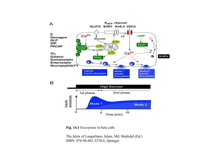
Fig. 14.1 Exocytosis in beta cells The Islets of Langerhans, Islam, Md. Shahidul (Ed.) ISBN: 978-90-481-3270-6, Springer. Fig. 14.2 Analytical methods used to study exocytosis and endocytosis. Adapted from [62] with permission from Elsevier

E N D
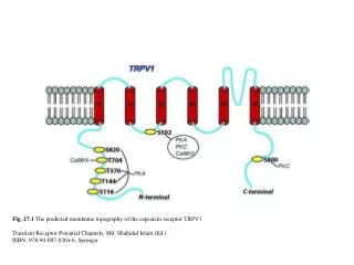
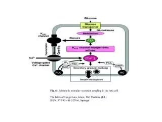
Fig. 14.1 Exocytosis in beta cells The Islets of Langerhans, Islam, Md. Shahidul (Ed.) ISBN: 978-90-481-3270-6, Springer
Fig. 14.2 Analyticalmethods used to study exocytosis and endocytosis. Adapted from [62] with permission from Elsevier The Islets of Langerhans, Islam, Md. Shahidul (Ed.) ISBN: 978-90-481-3270-6, Springer
Fig. 14.3 Different forms of compound exocytosis.Adapted from [61] with permission from Elsevier The Islets of Langerhans, Islam, Md. Shahidul (Ed.) ISBN: 978-90-481-3270-6, Springer
Fig. 14.4 Two-photon excitation imaging of exocytotic events in beta cells in mouse pancreatic islets. Adapted from [26] with permission from AAAS The Islets of Langerhans, Islam, Md. Shahidul (Ed.) ISBN: 978-90-481-3270-6, Springer
Fig. 14.5 Diameters of exocytotic vesicles and the fusion pore The Islets of Langerhans, Islam, Md. Shahidul (Ed.) ISBN: 978-90-481-3270-6, Springer
Fig. 14.6 Redistribution of SNAP-25 during sequential exocytosis.Adapted from [2] with permission from the Rockefeller University Press The Islets of Langerhans, Islam, Md. Shahidul (Ed.) ISBN: 978-90-481-3270-6, Springer
Fig. 14.7 Effects of cytosolic cAMP, ATP, and extracellular glucose on Ca2+ -induced exocytosis in betacells. (A–C) are adapted from [91] with permission from National Academy of Sciences, USA and (D–G) from [31] with permission from the Physiological Society The Islets of Langerhans, Islam, Md. Shahidul (Ed.) ISBN: 978-90-481-3270-6, Springer
Fig. 14.8 TEP imaging of exocytosis and endocytosis using FM1-43 in isolated beta cells.Adapted from [10] with permission from the Physiological Society The Islets of Langerhans, Islam, Md. Shahidul (Ed.) ISBN: 978-90-481-3270-6, Springer
Fig. 14.9 Ultrastructural identification of endocytotic vesicles in beta cells. Adapted from [10] with permission from the Physiological Society The Islets of Langerhans, Islam, Md. Shahidul (Ed.) ISBN: 978-90-481-3270-6, Springer
Fig. 14.10 Pharmacology of Ca2+-dependent exocytosis of SLMVs in beta cells. Adapted from [10] with permission from the Physiological Society The Islets of Langerhans, Islam, Md. Shahidul (Ed.) ISBN: 978-90-481-3270-6, Springer