Download
1 / 124
1.24k likes | 1.47k Views
Ventricular tachycardia in abnormal heart. Dr Ranjith MP Senior Resident Department of Cardiology Government Medical college Kozhikode. Ventricular tachycardia in abnormal heart OUTLINE. Coronary Artery Disease Dilated Cardiomyopathy Bundle Branch Re-entry Ventricular Tachycardia

E N D
Ventricular tachycardia in abnormal heart Dr Ranjith MP Senior Resident Department of Cardiology Government Medical college Kozhikode
Ventricular tachycardia in abnormal heartOUTLINE • Coronary Artery Disease • Dilated Cardiomyopathy • Bundle Branch Re-entry Ventricular Tachycardia • Arrhythmogenic Right Ventricular Cardiomyopathies • Hypertrophic Cardiomyopathy • After Surgery for Congenital Heart Disease
Ventricular tachycardia in abnormal heartOUTLINE • Coronary Artery Disease • Dilated Cardiomyopathy • Bundle Branch Re-entry Ventricular Tachycardia • Arrhythmogenic Right Ventricular Cardiomyopathies • Hypertrophic Cardiomyopathy • After Surgery for Congenital Heart Disease
VT in Patients with CAD • Healed MI is the most frequent clinical setting for the development of sustained VT • The first episode of VT can occur years after infarct healing • Clinical presentation- tolerated sustained VT to SCD • Incidence reduced from 3% to 1%
VT in Patients with CADMechanism of VT in a/c ischemia • Focal activation by abnormal automaticity in the ischemic border zone • a/c ischemia activates KATP channels causing an increase in extracellular K along with acidosis and hypoxia in the cardiac muscle • Minor increases in extracellular K depolarize the myocardiocyte’s RMP , which can increase tissue excitability in early phases of ischemia
VT in Patients with CADMechanism of VT in a/c ischemia • Focal discharge by Ca overload & triggered activity in the form of delayed or early after-depolarizations - not been proven experimentally
VT in Patients with CADMechanism • Reentry is the mechanism underlying the VT associated with healed or healing MI (>95%) • The ability to reproducibly initiate and terminate VT with programmed ventricular extrastimuli- the sine qua non of reentry • Induction of VT in coronary disease is stimulation site specific • An inverse relationship of the extrastimulus coupling interval to the onset of the first tachycardia beat is observed in many VTs
VT in Patients with CADPathophysiologic Substrate • Important determinants of arrhythmia risk after MI • The extent of myocardial necrosis • Presence of septal involvement • Degree of left ventricular dysfunction • Anatomic substrate – extensive scar • VT consistently arises from surviving myocytes within extensive areas of infarction • Conduction is slow & discontinuous, owing to fibrosis and abnormalities in gap junction distribution & function
VT in Patients with CADPathophysiologic Substrate • Electrophysiologic substrate for VT develops in the first 2 weeks after MI - remain indefinitely • Triggers – Acute ischemia – Surges in the autonomic tone – Heart failure • Once sustained monomorphic VT occurs, risk continues indefinitely, even if acute ischemia & heart failure are adequately controlled Josep Brugada et al JACC Vol. 37, No. 2, 2001:529–33
VT in Patients with CADPathophysiologic Substrate • Reentry – macro/ micro reentry • Repolarization of individual myocardial cells not homogenous. Some cells excitable, some refractory
VT in Patients with CADPathophysiologic Substrate Sinus rhythm mapping in a patient with VT in the setting of extensive healed anterior infarction. The map is color-coded to represent bipolar electrogram voltage: red (representing dense scar) denotes = 0.5 mV, purple = 1.5 mV, and the intervening colors represent voltage values in between. Multiple inducible VTs of varied morphology were localized to circuits within the scar
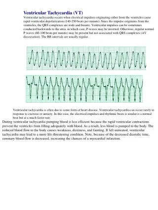
VT in Patients with CADECG findings • In the setting of an old MI, the ECG during VT is affected by • The size of infarction • The region of infarction • The region within the scar where the circuit is located • The proximity to the His-Purkinje system • The influence of concomitant pharmacological agents
VT in Patients with CADECG evidence of MI • Presence of Q waves (qR, QR or Qr) in related leads • Notched or wide QRS complexes • Low QRS voltage • Multiple ventricular tachycardia morphologies • Paroxysmal sustained episodes
VT in Patients with CADIdentifying the VT origin • Surface ECG tends to locate the reentry circuit exit rather than the VT origin • Location should be defined in 3 axes: • septal vs lateral walls • superior vs inferior walls • apical vs basal regions • Bundle branch block patterns -sequence of ventricular activation
VT in Patients with CADIdentifying the VT origin • Lateral wall VT • RBBB pattern • Wider QRS complexes • Septal VT • LBBB pattern • Narrower QRS complexes
VT in Patients with CADIdentifying the VT origin • The QRS axis in inferior leads indicates the sequence of activation between the superior and inferior walls • Inferior MI • Superior axis (80%) • Anterior MI • Superior axis (55% ) • Inferior axis (45%)
VT in Patients with CADIdentifying the VT origin • Predominant polarity of QRS complexes in precordial leads can help discriminate between VTs from the basal or the apical regions • VT from the apex • Negative concordant R progression • VT from the basal • Positive concordant R progression
VT in Patients with CADClinical Presentation & Management • The key determinant of hemodynamic tolerance • Tachycardia rate • Left ventricular function • Development of ischemia, and mitral insufficiency • Hemodynamic collapse – cardioversion • Intravenous procainamide, sotalol, and amiodarone have been demonstrated to have superior efficacy
VT in Patients with CADLong-Term Management • Goal of long-term therapy- prevention of SCD & recurrence of symptoms • Asymptomatic NSVT with NLVEF- no treatment • Symptomatic NSVT in pts with NLVEF- betablockers • Cardiac arrest survivors / SUS VT in ↓LVEF- ICD • Primary pvt - ICD > Amiod- pvt of SCD
VT in Patients with CADLong-Term Management • subendocardial resection of arrhythmogenic focus • Cryoablation • Laser vaporization • Photocoagulation
Primary Prevention of SCD in absence of Ventricular Arrhythmias
I I I I I I I I I IIa IIa IIa IIa IIa IIa IIa IIa IIa IIa IIa IIa IIb IIb IIb IIb IIb IIb IIb IIb IIb IIb IIb IIb III III III III III III III III III III III III I I I I I I I I I IIa IIa IIa IIa IIa IIa IIa IIa IIa IIa IIa IIa IIb IIb IIb IIb IIb IIb IIb IIb IIb IIb IIb IIb III III III III III III III III III III III III I I I I I I I I I IIa IIa IIa IIa IIa IIa IIa IIa IIa IIa IIa IIa IIb IIb IIb IIb IIb IIb IIb IIb IIb IIb IIb IIb III III III III III III III III III III III III A A A Current recommendations for ICD implantation in the setting of CAD • Patients resuscitated from VF when coronary revascularization is not possible, and there is evidence of prior MI and significant LV dysfunction • LV dysfunction due to MI who present with hemodynamically unstable VT • Primary prevention - LV dysfunction due to prior MI who are at least 40 days post-MI and have an LVEF 30%-40% & NYHA II or III
IIa IIa IIa IIa IIa IIa IIa IIa IIb IIb IIb IIb IIb IIb IIb IIb III III III III III III III III I I I I I I IIa IIa IIa IIa IIa IIa IIa IIa IIb IIb IIb IIb IIb IIb IIb IIb III III III III III III III III I I I I I I IIa IIa IIa IIa IIa IIa IIa IIa IIb IIb IIb IIb IIb IIb IIb IIb III III III III III III III III I I I I I I \ Current recommendations for ICD implantation in the setting of CAD • Primary prevention ICD is reasonable in patients with LV dysfunction due to prior MI who are at least 40 days post-MI, and have an LVEF 30%-35% & NYHA I • ICD implantation is reasonable in patients with post-MI with normal LV function and recurrent VT B C
Ventricular tachycardia in abnormal heartOUTLINE • Coronary Artery Disease • Dilated Cardiomyopathy • Bundle Branch Re-entry Ventricular Tachycardia • Arrhythmogenic Right Ventricular Cardiomyopathies • Hypertrophic Cardiomyopathy • After Surgery for Congenital Heart Disease
Ventricular tachycardia in abnormal heartOUTLINE • Coronary Artery Disease • Dilated Cardiomyopathy • Bundle Branch Re-entry Ventricular Tachycardia • Arrhythmogenic Right Ventricular Cardiomyopathies • Hypertrophic Cardiomyopathy • After Surgery for Congenital Heart Disease
VT in Patients with DCM • DCM has a propensity to the development of ventricular arrhythmias and sudden death • Incidence of DCM - 4 to 8 cases per 100,000 population • Incidence of VT – 50-60% DCM, resp for 8-50% deaths • Genetics - Relationship between individual genotypes and arrhythmogenicity is poorly understood
VT in Patients with DCMArrhythmogenesis • Multiple factors responsible for VT in DCM
VT in Patients with DCMArrhythmogenesis • Myocardial fibrosis/scar - may act as sites for reentry • At autopsy, extensive subendocardial scarring in the LV in 33% & multiple patchy areas of replacement fibrosis in 57% • Sustained stretch-induced shortening of refractory period and AP duration, predisposing to reentry • Short, pulsatile, stretch-induced after depolarizations
VT in Patients with DCMArrhythmogenesis • Diastolic Ca overload caused by decreased sacrcoplasmic reticulum Ca2+–adenosine triphosphatase pump • Afterdepolarizations induced by increased Na+-Ca2+ exchanger activity • Hypokalemia, hypomagnesemia (often related to diuretic use) • Increased circulating catecholamines
VT in Patients with DCMArrhythmogenesis • Increased sympathetic tone • Purkinje system conduction delay • Increased endocardial surface area in dilated atrium or ventricle • Drugs (antiarrhythmics, digoxin, sympathomimetic)
VT in Patients with DCMMechanisms • Macro reentry - dominant mechanism • Bundle branch reentry ventricular tachycardia (BBRVT) is the most characteristic • BBRVT - Responsible for VT in up to 41% of DCM • Macro-reentrant circuit involving the His-Purkinje system, usually with antegrade conduction over the RBB and retrograde conduction over the LBB
VT in Patients with DCMPredictors of Mortality &Ventricular Arrhythmias • Severity of LV dysfunction - most powerful predictor • ISCD is significantly greater in patients with syncope • Laboratory values - low serum sodium and increased plasma norepinephrine, renin, and ANP,BNP • LBBB & of first- and second-degree AV block has been associated with poor outcome
VT in Patients with DCMPredictors of Mortality &Ventricular Arrhythmias • Vesnarinone Trial (VEST) showed a significant association between the degree of QRS prolongation and mortality Gottipaty V, et al.J Am CollCardiol 33:145A, 1999
VT in Patients with DCMTreatment • ACEI – reduction in SCD due to VT (37% vs 46%) • new VT developed less frequent at 1,2 yrs in enalapril group (VHeFT-II trial)
VT in Patients with DCM Treatment • Amiodarone • Used only on specific arrhythmic indications • Reduces ICD shock frequency , without worsening heart failure (SCDHeFT) • Implantable Cardioverter-Defibrillators • AMIOVIRT-No difference in mortality ( amio vs ICD) • SCD-HeFT - Significant reduction in total mortality in ICD group • Catheter ablation
VT in Patients with DCM Treatment • Biventricular pacing- severe drug refractory heart failure , in elderly • Improve systolic function by shortening the duration of mechanical systole and increasing dP/dt • Improve diastolic function by prolonging diastolic filling time • Reduce presystolic MR by earlier activation of the lateral papillary muscle without the adverse effect on the sympathetic nervous system seen with inotropic agents
Ventricular tachycardia in abnormal heartOUTLINE • Coronary Artery Disease • Dilated Cardiomyopathy • Bundle Branch Re-entry Ventricular Tachycardia • Arrhythmogenic Right Ventricular Cardiomyopathies • Hypertrophic Cardiomyopathy • After Surgery for Congenital Heart Disease
Ventricular tachycardia in abnormal heartOUTLINE • Coronary Artery Disease • Dilated Cardiomyopathy • Bundle Branch Re-entry Ventricular Tachycardia • Arrhythmogenic Right Ventricular Cardiomyopathies • Hypertrophic Cardiomyopathy • After Surgery for Congenital Heart Disease
Bundle Branch Re-entry VT • Commonly occurs in disease with severe LV dysfunction like DCM & conduction abnormalities in the HPS • BBR VT may also be seen in: • Myotonic dystrophy • Hypertrophic cardiomyopathy • Ebstein anomaly • Following valvular surgery • Proarrhythmia due to Na channel blockers • Presyncope, syncope or sudden death - VT with fast rates > 200 bpm
Bundle Branch Re-entry VTMechanisms • Macro re-entrant circuit employing • His Bundle • Both bundle branches • Ramifications of left bundle • Transeptal myocardium
Bundle Branch Re-entry VTMechanisms • May present with LBBB or RBBB morphology depending on the antegrade conduction • LBBB morphology is common BBR –LBBB: - antegrade direction -RB & reterograde LB BBR –RBBB:- antegrade direction-LB & reterograde RB
Bundle Branch Re-entry VTMechanisms • Surface ECG in sinus rhythm - non-specific or typical bundle branch block patterns with prolonged QRS duration • Total interruption of conduction in one of the BB would theoretically prevent occurrence of reentry • Can occur in patients with relatively narrow QRS complex - functional conduction delay
Bundle Branch Re-entry VT Electrophysiologic features • During tachy QRS morphology is commonly LBBB type • His electrograms precede each V • HV interval during tachycardia > HV in baseline • Changes in V–V interval follow the changes in H–H • Delay in HPS conduction facilitates induction