Download
1 / 51
510 likes | 648 Views
Gray and White Matter Abnormalities in ADHD: An Optimized Voxel-Based Morphometry Study. Carmona S. (1), Bielsa A (3) ,Trèmols V. (2), Soliva J.C. (4), Rovira M. (4), Tomàs J. (3), Bulbena, A (1). Raheb, C,. (3), Gispert J. D. (5) , Tobeña A. (1), Vilarroya O. (1)

E N D
Gray and White Matter Abnormalities in ADHD:An Optimized Voxel-Based Morphometry Study Carmona S. (1), Bielsa A (3) ,Trèmols V. (2),Soliva J.C. (4), Rovira M. (4), Tomàs J. (3), Bulbena, A (1). Raheb, C,. (3), Gispert J. D. (5) , Tobeña A. (1), Vilarroya O. (1) Dep. Psiq. Med. Leg. Univ. Autonoma Barcelona, Spain 1, USP Inst. Univ. Dexeus, Barcelona, Spain 2, Serv. Paidopsiq. Hosp. Vall d’Hebron, Barcelona, Spain 3, CRC Corporacio Sanitaria, Barcelona, Spain4, Institut d’Alta Tecnologia – Parc de Recerca Biomèdica de Barcelona, Spain 5.
INTRODUCCIÓN • TDAH: • DESATENCIÓN • HIPERACTIVIDAD • IMPULSIVIDAD Interfieren en el funcionamiento del niño en diferentes ambientes Anterior a los 7 años
Perfil: • Dificultad para focalizar y mantener la atención. Déficits de WM. • Dificultad para inhibir impulsos. Pobres habilidades de planificación. • Dificultad para controlar el movimientos. Pocas habilidades motrices (fina y gruesa). • Déficits emocionales. • Déficits en las relaciones sociales.
Substrato Neural: Implicación del circuito FRONTO-ESTRIATAL Derecho con modulación Cerebelar en la sintomatología del TDAH. (Barkley 1998, Giedd 2001) Otros circuitos implicados??? “…circuits originating in supplementary and other premotor areas.” (Mostofsky 2002) INTRODUCCIÓN
INTRODUCCIÓN • Anormalidades estructurales más replicadas en ADHD • Globales: • REDUCCIÓN: • VOLUMEN TOTAL CEREBRAL (3.2%-8%) • VOLUMEN DE SUSTANCIA GRIS • VOLUMEN DE SUSTANCIA BLANCA • Regionales: • Sustancia Gris: • DISMINUCIÓN DEL CEREBELO (Posterior Lobes, nº VIII-X/ VI-VII) • Anormalidades en el CórtexPrefrontal (Derecho/Izquierdo/Asimetría anormal) • Anormalidades enelCaudado (Derecho/Izquierdo/Asimetría anormal) • Sustancia Blanca: • Reducción de SB en ÁreasFrontales Derechas • Redución del Rostrum del Cuerpo Calloso (Reviews: Eliez 2000, Giedd 2001,Leo 2003, Castellanos 2004 )
INTRODUCCIÓN • Otros hallazgos estructurales: • Globales: • No diferencias en el Volumen Total Cerebral (Filipek 1997, Lyoo 1996) • No diferencias en el Volumen de Sustancia Gris(Sowell, 2003. ) • Regional: • Sustancia Gris: • Reducción del Córtex Premotor(Mostofsky, 2002) • Reducción del Córtex Cingulado Medial (Filipek, 1997) • Reducción del Córtex Cingulado Posterior(Overmeyer, 2001) • Sustancia Blanca: • Reducción de SB predominantemente en el Hemisferio Izquierdo (Overmeyer, 2001; Mostofsky,2002) • Reducción del Splenium del Cuerpo Calloso(Semrud-Clikeman 1994)
INTRODUCCIÓN • La mayoría de estos estudios utilizan análisis volumétricos manuales, delimitando Regiones de Interés (ROI) previamente determinadas. • Optimized Voxel-Based Morphometry es un procedimiento automático que permite analizar el cerebro en su totalidad. (procesado automático de las imágenes voxel a voxel)
INTRODUCCIÓN: OBJETIVOS • Objetivo I: Diferencias Globales • Comparar el Volumen Total Cerebral (VTC), los Volúmenes de Sustancia Gris (SG), Sustancia Blanca (SB) y Líquido Céfalo-Raquídeo (LCR) entre ADHD y Controles. • Objetivo II: Diferencias Regionales • Analizar, en todo el cerebro, posibles diferencias regionales de SG y SB entre niños con ADHD y Controles.
INTRODUCCIÓN: HIPÓTESIS • El grupo de niños con ADHD tendrán menor: • VOLUMEN TOTAL CEREBRAL • Volumen Total de SG • Volumen Total de SB • Las diferencias estarán localizadas en: • SG: • Cerebelo • Córtex Prefrontal • Núcleo Caudado • SB: • Cuerpo Calloso.
MÉTODOS: Participantes • DESCRIPTORES Comb.: Combined Subtipe; I-H: Impulsive-Hyperactive subtipe; Inatt: Inattention subtipe. R: Right-handed, L: Left-handed, C.D.:Cross Dominance mg/kg: Methilphenidate miligrams per kilogram
MÉTODOS: Participantes • INCLUSION CRITERIA
MÉTODOS: Adquisición de las Imágenes • Las imágenes se obtuvieron en un scanner (GE Signa General Electric) de 1.5T. Se adquirió una imagen 3D SPGR potenciadas en T1 para cada sujeto. • Parámetros de adquisición: • TE =4.2 ms • TR = 13.2 ms • Flip angle = 15º • Slice plane = axial • Matrix size = 256 x 256 • Slice thickness = 2 mm.
MÉTODOS: Pre-procesado de Imágenes • Inspección visual de las imágenes en formato Analyze para asegurar su cualidad (ausencia de movimiento o artefactos de adquisición, orientación correcta en el eje AC-PC….). • Creación de plantillas (global, SG, SB y LCR) específicas y derivadas a partir de la muestra de sujetos. • Aplicación del procedimiento Optimized-voxel-based-morphometry(Good et al.2000, 2001)
MÉTODOS: Procesado de Imágenes • Optimized Voxel-Based Morphometric Approach: • Segmentación de la imagen 3d en SG,SB y LCR de acuerdo con la plantilla de todo el cerebro previamente creada. • Cálculo del Volumen de SG, SB, LCR y Total. • Normalización de la imagen de SG y SB a basada en la plantilla correspondiente (Gris o Blanca en función del objetivo del análisis). • Segmentación de la imagen previamente normalizada. • Modulación de la imagen normalizada y segmentada con el determinante Jacobiano derivado de la deformación al normalizar la imagen. • Suavizado de las imágenes moduladas con un Gaussian kernel de 12 x 12 x 12 mm FWHM . VSG = 759,26 cm3 VSB = 409,42 cm3 VLCR = 278,31 cm3 VTC = 1446,99 cm3
MÉTODOS: Análisis Estadístico • Volumétricas Globales, (VTC, VSG, VSB, VLCR): • Prueba T-Test comparación de medias para muestras independientes. • SPSS • Análisis morfométrico regional: • Prueba T-Test de comparación de medias para muestras independientes. • SPM2 • Dos contrastes (Controles>ADHD; ADHD>Controles) para cada una de las comparaciones (SG; SB). El umbral de significación fué P< 0.01 (FDR) y sólo se tuvieron en cuenta aquellas diferencias superiores a los 10 vóxeles contiguos.
RESULTADOS • T-test de las medidas Volumétricas Globales: • Los niños con ADHD presentan una reducción de alrededor del 5.4% en el VTC y del 5.2% en el VSG. • No se observaron diferencias significativas en otras medidas.
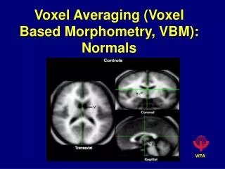
RESULTADOS • Diferencias Regionales de SG: p< 0.01 FDR-corrected • No diferencias en SB
RESULTADOS : Reducciones en el Volumen de SG en niños ADHD • Lóbulo Frontal • Córtex Premotor • Giro Precentral Derecho • Córtex Orbitofrontal • Circunvolución Recta Derecha • Córtex Dorsolateral • Opérculo Rolándico Izquierdo • Fronto-Parietal • Córtex Somatosensorial, Motor y Premotor • Áreas perirrolándicas Izquierdas • Lóbulo Temporal • Giro Temporal Medio Bilateral • Córtex Cingulado • Córtex Cingulado Posterior y Medial Izquierdo • Formación Hipocampal • Giro Parahipocampal Izquierdo • Cerebelo • Lóbulos Posteriores (Crus 1 y 2) bilateralmente
DISCUSIÓN • Diferencias Globales: • Los niños con ADHD presentan un menor Volumen Cerebral Total. • La reducción oscila entre 133.35 and 20.37 mililitros cúbicos. • En promedio una reducción del 5.4% • Los niños con ADHD presentan un menor Volumen de Sustancia Gris. • La reducción oscila entre 67.38 a 12.93 mililitros cúbicos. • En promedio una reducción del 5.2% • No se observaron diferencias en SB ni LCR.
DISCUSIÓN • AREAS FRONTALES: • Córtex Dorsolateral • Córtex Orbitofrontal Dificultad para focalizar y manterner la Atención. Déficits de WM Dificultad para inhibir impulsos. Pobres habilidades de planificación • CIRCUITOS MOTORES: • Areas Motoras y Premotoras • Cerebelo • CIRCUITOS LÍMBICOS: • Córtex Cingulado M/P • Formación hipocampo-amig Dificultades planificación y control del movimiento Pocas habilidades motrices Escaso control Emocional Motivación (distractibilidad)
CONCLUSIÓN • Las alteraciones que se observan en niños con ADHD no son únicamente debidas a disfunciones en el Circuito Fronto-Estriado-Cerebelar Derecho. • Implicación de otros circuitos (Circuitos Motores, Circuitos Límbicos…) especialmente sensibles a alteraciones durante el neurodesarrollo (Genéticos, obstétricos, Perinatales, ambientales…), que darían lugar a los diferentes síntomas.
INTRODUCCIÓN • Otros hallazgos estructurales: • Globales: • No diferencias en el Volumen Total Cerebral (Filipek 1997, Lyoo 1996) • No diferencias en el Volumen de Sustancia Gris(Sowell, 2003. ) • Regional: • Sustancia Gris: • Reducción del Córtex Premotor(Mostofsky, 2002) • Reducción del Córtex Cingulado Medial (Filipek, 1997) • Reducción del Córtex Cingulado Posterior(Overmeyer, 2001) • Reducción del Gobus Pallidus (Izquierdo/ Derecho) (Aylward 1996, Castellanos 1996) • Sustancia Blanca: • Reducción de SB predominantemente en el Hemisferio Izquierdo (Overmeyer, 2001; Mostofsky,2002) • Reducción del Splenium del Cuerpo Calloso(Semrud-Clikeman 1994)
DISCUSIÓN • Regional differences: • ADHD reviews: (Eliez 2000, Giedd2001,Leo 2003, Castellanos 2004 ) • GM • Reduced Cerebellum • Abnormalities in Prefrontal Córtex • Abnormalities in Caudate Nucleus (Castellanos 2004) Bilateral Reduction of Cerebellum * GM reduction of : Left Dorsolateral Córtex Right Orbitofrontal Córtex No differences due to the wide interval of age of our sample (6 to 16) Age affect specially differences Caudate Nucleus (Castellanos 2002)
DISCUSIÓN • Reducción: • Cerebelo: Déficits de coordinación motriz. • C.P.F.: • DL: WM • OF: Impulsividad • ADHD reviews: (Eliez 2000, Giedd2001,Leo 2003, Castellanos 2004 ) • GM • Reduced Cerebellum • Abnormalities in Prefrontal Córtex • Abnormalities in Caudate Nucleus
CONCLUSIÓN • Circuits: • Fronto-Striatal Circuits: • Motor Circuits: • Left Sensorimotor and Supplementary Motor Área (perirolandic Córtex) reductions • Bilateral Cerebellum reductions • Limbic Circuits: • Middle/Posterior and Cingulate Córtex reduction • Left Parahipocampal Gyrus reduction Consistent with motor abnormalities in ADHD Consistent with motovational-distractibility components of attention processes
DISCUSIÓN • Replicated findings: • Reduced Premotor Córtex: • Filipek 1997; Smaller volumes in Right Supplementary Motor Area (SMA) • Mostofsky 2001; Premotor GM reductions Bilaterally • fMRI: SMA seems to be important in response inhibition and present hypoactivation in ADHD patients during inhibition task (Krieger 2003, Tam 2004) • Reduced Middle/Posterior Cingulate Córtex: • Filipek 1997; Smaller volumes in Middle Cingulate Córtex • Overmeyer 2001;Smaller Posterior Cingulate Córtex (Hyperkinetics) • fMRI: Small et al 2003: “The funtion of the posterior cingulate Córtex apears to entail an inhibition of parietal cortices, probably for preventing distractibility” • Other findings: • Reduced Bilateral Middle Temporal Gyrus: • Temporal Lobe GM was the most significantly reduction of all GM regions studied by Castellanos in 2002. • fMRI: Shafritz 2004 reported that ADHD recruited significantily less Middle Temporal Gyrus. “MTG mediates attentional processing for verbal stimuly and maybe particulary important when attending to auditory information” • Reduced Left Parahippocampal Gyrus: • Low birth weight children with ADHD have smaller bilateral hippocampal formation than low birth weight children without ADHD (Abernety 2002)
DISCUSSION • Neurodevelopmental Disorders: • Most of the regions of reduced GM in ADHD have been found also in other neurodevelopmental disorders. For example: • Preterm children: • Decreased Total Cerebral Volume • Reduced Cerebellum • Reduced Left parietal Lobe • Reduced Sensorimotor Córtex • Reduced Middle Temporal Areas • Reduced Hippocampus • There is also a hight prevalence of comorbility or symptoms overlaping between neurodevelpomental disorders. For example: • ADHD: • Conduct Disorder: Emotional and social impulsive behaviour. • Tourette Disorder: Motor control defficits and social and emotion disinhibition. • Obsessive Compulsive Disorder: Failure to inhibit impulses. • … • Cerebral regions specially sensitive to be damage Bradley 2000,Peterson 2000, Isaacs 2000, 2001,Allin 2001, Abernethy 2002
DISCUSSION • Regional differences: • P<0.01 FDRcorrected. • Left Dorsolateral Prefrontal Córtex: • Right Orbitofrontal Córtex: • Bilateral Cerebellum Posterior Lobes : • Those findings support previous findings about anatomical abnormalities in ADHD. • Prefrontal Regions had been related to impulsive behaviour, specially those involving Right Orbitofrontal areas. Other prefrontal areas such as those located in Dorsolateral Córtex seem to be more related to defficits in Working Memory or other impariments in executive functions that can be found in ADHD children. • As Castellanos says in 2004, Cerebellum defficit is the most consistent finding across MRI ADHD literature, and also is one of the areas that remains significant after FWE correction. Most of the studies found cerebellar defficits located in Posterior Lobes, nº VIII-X/ VI-VII. We found reductions only in Lateral Posterior Lobules (Crus 1 and 2), surely this discrepance is because our images do not scan the whole cerebellar lobes and we don’t have information about lobes from VI to X.
DISCUSSION • Regional differences: • Replicate previous finddings: • Reduced Cerebellum Bilaterally • Reduced Prefrontal Córtex: • Left Dorsolateral Córtex • Right Orbitofrontal • Reduced Premotor Córtex (Filipek 1997, Mostofsky 2002) • Reduced Middle/Posterior Cingulate Córtex ((Filipek 1997, Mostofsky 2001) • Other findings: • Bilateral reduction of Middle Temporal Gyrus • Left Parahippocampal Gyrus Consistent findings (Reviews: Eliez 2000, Giedd 2001, Leo 2003, Castellanos 2004 )
Left Parahippocampal Gyrus: • Left Middle Posterior Cingulate Gyrus: • Bilateral (predominantly left-sided) Premotor Córtex: • Bilateral Middle Temporal Gyrus:
DISCUSSION • Regional differences: • P<0.05 FDRcorr. • Generalized GM reducctions involving • Fronto-Parieto-Temporo-Occipital regions Bilaterally. (Castellanos 2002, Sowell 2003) • Different Prefrontal Regions, specially Right Orbitofrontal. • Right Caudate (Castellanos 1994) • Amigdala Bilaterally. (Castellanos 2001) • Cerebellum Bilaterally Replicated findings (Eliez, Giedd, Leo and Castellanos’s reviews)
RESULTS • REGIONAL GM DIFFERENCES: p< 0.05 FDR-corrected • NO WM DIFFERENCES
RESULTS • LESS GRAY MATTER VOLUME • IN ADHD GROUP • P< 0.05 FDR-corrected
Fronto-Parieto-Temporo-Occipital regions Bilaterally. Predominantly Left Fronto-Parieto-Temporal and Right Orbitofrontal. • Right Caudate (Castellanos 1994) • Cerebellum Bilaterally. • Subgenual, Middle and Posterior Cingulate Cyrus Bilaterally. • Bilateral Parahippocampal Formation and Amigdala Nuclei
P<0.05 FDR-corrected P<0.01 FDR-corrected RESULTS: GM volume reducctions in ADHD group
RESULTS: GM volume reducctions in ADHD group • Frontal Lobe • Premotor Córtex • Right Precentral Gyrus (BA 6) • Left Precentral Gyrus (BA 6) • Left Postcentral Gyrus (BA 4) • Left Postcentral Gyrus (BA 4) • Orbitofrontal Córtex • Right Rectal Gyrus (BA 11) • Dorsolateral Córtex • Left Rolandic Oper (BA 48)
RESULTS: GM volume reducctions in ADHD group • Temporal Lobe • Right Middle Temporal Gyrus (BA 21) • Left Middle Temporal Gyrus (BA 22) • Cingulate Córtex • Left Middle Cingulate Gyrus (BA 23) • Left Middle Cingulate Gyrus • Left Posterior Cingulate Gyrus (26) • Parahippocampal Formation • Left Parahippocampal Gyrus (BA 36)
Cerebellum • Left Posterior Lobe
Funtional Neuroimage • Frontal Parietal Regions Failed to be recruited • Anterior Cingulate Córtex • Orbitofrontal Córtex
Total Brain Volume • The study with a bigger n : 3.2% reducced. (Castellanos 2002): • Unmedicated ADHD had smaller Total Brain Volume in comparison with Control Subjects
INTRODUCCTION • MORE REPLICATED MRI FINDINGS IN ADHD (Reviews: Eliez 2000, Giedd 2001,Leo 2003, Castellanos 2004 ) • TOTAL BRAIN VOLUM (3.2% TO 8%) • Specific regions: • GM structures: • Prefrontal Córtex • Basal Ganglia: Caudate (also Putament and Globus Pallidus) • CEREBELLUM (Posterior Lobes, nº) • WM Structures: • Corpus Callosum: • Rostral • Splenium
INTRODUCCTION • MORE REPLICATED MRI FINDINGS IN ADHD (Reviews: Eliez 2000, Giedd 2001,Leo 2003, Castellanos 2004 ) • TOTAL BRAIN VOLUM (3.2% TO 8%) • Specific regions: • GM structures: • Prefrontal Córtex • Basal Ganglia: Caudate (also Putament and Globus Pallidus) • CEREBELLUM (Posterior Lobes, nº) • WM Structures: • Corpus Callosum: • Rostral • Splenium
P<0.05 FDR-corrected P<0.01 FDR-corrected RESULTS: GM volume reducctions in ADHD group