Download
1 / 1
10 likes | 146 Views
Cancer Metastasis Study with Animal Extracts. Michael Lau , Priya Garigipati , Cristian Hernandez, Akash Sharma, Mahrukh Yasin , Stephen Ahn , Smitha Rao, Victor Lin, Uday Tata, J.-C. Chiao Electrical Engineering Summer REU, The University of Texas at Arlington, Arlington, Texas 76019.

E N D
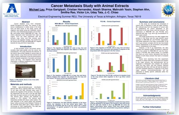
Cancer Metastasis Study with Animal Extracts Michael Lau, PriyaGarigipati, CristianHernandez, Akash Sharma, MahrukhYasin, Stephen Ahn, Smitha Rao, Victor Lin, Uday Tata, J.-C. Chiao Electrical Engineering Summer REU, The University of Texas at Arlington, Arlington, Texas 76019 Abstract Cancer fatalities occur from metastatic growth throughout the body. The causes of why cancer metastasizes to certain areas in the body have not been found. In this study, cancer cell migration was tested using two metastatic cancer cell lines, lung metastasized prostate cancer (PC3-ML) and breast cancer (MDA-MB 231) cells. The chemo attractants were harvested from male rat organs. From the initial experiment with the extracts, lung, femur and liver extracts were chosen for the migration studies. The results thus far confirm that PC3-ML and MDA-MB 231 migrate towards the lung and femur respectively. Introduction In both prostate cancer and breast cancer, mortality rates raise greatly once the cancer has metastasized. This study observes the behavior of the cancer cells as they migrate to various male rat organ extracts. Lung, femur and liver extracts were used as chemo attractants to test the phenomenon of the metastatic properties of prostate and breast cancer cell lines. The migration study observes chemotaxis in different suspensions of extract with 2, 4 and 8 µg concentrations. Summary and conclusions In this study, we monitored the metastasis of cancer cells in response to male rat organ extracts. We collected data from the migration of cancer cells by establishing the chemo attractants of the experiment to be lung, femur, and liver extracts. The chemotaxis of MDA-MB 231 and PC3-ML were observed with 2µg, 4 µg, and 8 µg concentrations of each organ extract. The trends produced from the data suggest that proteins in the femur and the lung extracts cause migration in MDA-MB 231 and PC3-ML respectively. The trends observed in the lower concentration (2 µg and 4 µg) extracts correlate with the fact that breast cancer metastasizes to the bone/femur and prostate cancer metastasizes to the lungs). However, in the 8 µg experiments, the data indicates that migration greatly increased in response to the liver extract for MDA-MB 231 and the femur extract in PC3-ML with similar trends. It is possible that there are some chemo attractants that may be present in both these extracts that lead to increased migration. Future work stemming from this experiment would be to assess what kind of proteins are present in the organ extracts. Gel electrophoresis will be employed to distinguish and compare the proteins within the lung, femur, and liver extracts. Identical experiments with other cell lines will also be tested to see their metastatic behavior in response to the same organ extracts. Immunohistochemistry will be performed to identify specific antibodies within the cells themselves. Results MDA-MB 231 – Extract Experiment PC3-ML – Extract Experiment Introduction Day 1 Day 5 Figure 5: The migration of PC3-ML cells to lung, liver and femur extracts displays that chemotaxis was greatest in response to the lung extract with mild response to the liver extract. Figure 2: The migration of MDA-MB 231 cells to lung, liver and femur extracts shows that migration was highest in response to the femur extract. Materials and methods PDMS (poly-dimethylsiloxane) microfluidic devices with 100 µL capacity wells and 1 mm long, 10 µm wide and high channels were used to observe the migration of the cell lines. The setup of the experiment has the cells in the left side wells with male rat organ extracts on the right side wells. The devices were cleaned and sterilized by soaking in 70% ethanol. The devices were temporarily bonded with tissue culture plates. The cells attached to the culture plates and standard tissue culture protocols were followed. Pictures of the channels were taken with a Nikon Eclipse TI microscope at 100X magnification. The data was processed using AxioVision Rel. 4.8. Figure 6: The data show that PC3-ML continues its migration trend to the lung extract while migration to the 4 µg femur extract increases. Figure 3: The migration of MDA-MB 231 to lung, liver and femur extracts displays that migration was absent from the lung and liver extract. The femur extract was still the highest. Literature cited “Demonstration of Cancer Cell Migration Using a Novel Microfluidic Device,” S. Rao, V. Lin, U. Tata, G. Raj, J.-T. Hsieh, K. Nguyen, and J.-C. Chiao, Journal of Nanotechnology for Engineering and Medicine, Vol. 1, No. 2, May 2010. Howlader N, Noone AM, Krapcho M, Neyman N, Aminou R, Altekruse SF, Kosary CL, Ruhl J, Tatalovich Z, Cho H, Mariotto A, Eisner MP, Lewis DR, Chen HS, FeuerEJ, Cronin KA (eds).SEER Cancer Statistics Review, 1975-2009 (Vintage 2009 Populations), National Cancer Institute. Bethesda, MD, http://seer.cancer.gov/csr/1975_2009_pops09/, based on November 2011 SEER data submission, posted to the SEER web site, 2012.. Figure 1: A Microfluidic device. A set of two wells constitutes a device. B F Acknowledgments We thank Dr. KambizAlavi and Mohammadreza Jahangir Moghadam for heading the REU Program. We also thank NSF and UTA for funding and hosting the REU program. NSF grant # EEC-1156801, REU Site: Research Experiences for undergraduates in Sensors and Applications at University of Texas at Arlington Figure 7: The disappearance of migration towards the lung extract could be attributed to cell death in the channel or that cells moved into the right well. Similar to the 8 µg MDA-MB 231 trend, the higher concentration femur extract could have similar protein concentration as the lower concentration lung extracts, leading to migration B D F Figure 4: Furthermore, the data indicates migration to the femur extract, but migration to the liver extract grew higher. This presents the possibility that the 8 µg liver extract has a similar concentration of specific proteins to the lower concentration femur extracts. Further Information Contact Michael Lau (mxl110330@utdallas.edu) or Smitha Rao (smitha@uta.edu).