Download
1 / 29
300 likes | 726 Views
University School “La Sapienza”, Rome Cutaneous and Venereal Deseases Department. Bowenoid Papulosis S. Pala, A. Spataro, I. Poleva. Introduction.

E N D
University School “La Sapienza”, Rome Cutaneous and Venereal Deseases Department Bowenoid Papulosis S. Pala, A. Spataro, I. Poleva
Introduction Bowenoid papulosis (BP) is an interesting pathology for its not well understood aethiology and variable clinical course: mainly benign, but sometimes associated with malignant invasive transformation (2.6%). BP may be considered to be a transitional state between a genital warts and Bowen disease. BP is manifested as papules that are induced virally by human papillomavirus (HPV) and demonstrates a distinctive histopathology (bowenoid dysplasia). Some sources argue that bowenoid papulosis, similar to condyloma acuminata, will resolve spontaneously, even after months or years. However, the behavior of HPV cannot be predicted histologically and local invasion seems to be more common in older adult patients and patients who are immunocompromised.
Case report • A 39 y.o. italian female patient HIV+ since 1987, reached the Sexually Transmitted Deseases Center of Policlinico Umberto I, University “La Sapienza” in Rome, for the 6 months history of painless papules in the vulval and perianal regions and a nodular lesion of the lower lip.
Physical examination • At the physical examination of the genital region coalescent, warty papules were observed. They had dark brown colour, 10- 15 mm in diameter. • There was a nodular lesion on the lower lip, elastic and pink in colour. • The rest of the physical examination was unremarcable • Biopsy of the nodular lesion of the lip and of the genital one was executed.
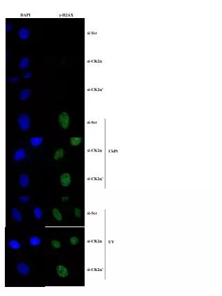
Histopathology The histopathological examination reveals: Topical acanthosis, koilocytosis, pleomorphous nucleous, apoptosis, numerous mitotic figures, irregular keratynocytes. These characteristics confirm the clinical suspicion of bowenoid papulosis.
University school “La Sapienza”, Rome DermoHystopatological service Prof. D. Innocenzi
University School “La Sapienza”, Rome DermoHystopatological sevice Prof. D. Innocenzi
HPV-DNA Research Research of HPV-DNA through specific PCR in both lesions reveals the presence of • HPV-DNA 32in the lower lip lesion • HPV-DNA 16in the genital lesion.
Historical notices • In 1970 Lloid describes this dermatosis for the first time like “multicenter pigmented Bowen’ s disease of the groin”. • Subsequently, in 1978, Wade introduced the term “Bowenoid Papulosis” in order to describe such pathology that is placed between Acuminate Condylomata and Bowen’ s disease. • In the same year Kimura describes a clinical case of viral pigmented papulosis of genitals.
Ethiopatogenesis • The BP ethiopathogenesis is not well understood still today. • HPV has been linked closely to BP. HPV is a very small DNA-virus with specific tropism for keratinocytes and the mucosas; • The more demonstrated in the lesions trough hybridization in situ viral genotypes are: HPV 16, HPV 18, HPV 31, HPV 32, HPV 33, HPV 39, HPV 42, HPV 48, HPV 51. • The infection by HPV 16, HPV 18, HPV 31 is an important factor of risk for the squamocellular carcinoma in situ, above all in HIV+ patients and in general in immunodepressed.
FREQUENCY BP lesions are related clinically to genital warts. They share the same age of onset and are transmitted sexually. Since BP lesions frequently are treated destructively as warts and without histopathologic examination, the true frequency of BP is unknown but is believed to be underestimated. A number of case reports associate BP with malignant invasive transformation (2.6%). All races are affected equally
Clinical manifestations • The young adults (average age 31 years), sexually active, hetero- and homosexual, are hit, with a light female predilection. • The lesions interest more frequently the genital, perianal and perineal areas, but in HIV+ patients also extragenital regions. • The morphology is very variable: warty, pink or brown or violet, well delimited macula-papulas, 2-30 mm in diameter; frequently asymptomatic, but it’ s possible also itch, erythema, hyperpigmentation and inflamation.
Histopathological features • The granular layer is thicken defined with hyperkeratosis, parakeratosis and dyskeratosis. • Cellular atypia: • Pleomorphic keratinocytes , hypercromatic and amassed magnified nucleous; some of them with numerous nuclei; • koilocytosis ( it’ s a cytoplasmatic vacuolation around the thickened nuclear chromatin ); • numerous mitotic figures and cells in metaphase; • if the lesion is coloured, melanin-laden cells; • often altered acrosyringia; • always integral acrotrichia.
University School “La Sapienza”, Rome DermoHystopatological service Prof. D. Innocenzi
Differential diagnosis The most important are Bowen’s disease and the Condylomatosis of the genitals.
Differential diagnosis • Bowen’ s disease The Bowen’ s disease differs for the following clinical and histopathological characteristics: • has its highest incidence in the older age groups; • arises mainly in sun-damaged areas; • usually single lesion; • it never disappears spontaneously; • erythematous, scaly patches or plaques that may become hyperkeratotic, crusted, fissured, or ulcerated; • full tickness dysplasia with loss of the normal maturation of its components • large pale keratinocytes with abundant ground glass cytoplasm, so-called pagetoid cells • integral acrosyringia; • altered acrotrichia;
Bowen’ s disease Courtesy of Arthur C. Hunley
Differential diagnosis • Acuminate condylomata • mainly occur on the genital, perianal and perineal regions; • flesh-coloured papular lesions; • cauliflower lesions; • hyperkeratosis, parakeratosis, acanthosis; • koilocytosis; • viral inclusions in the cytoplasm and the nucleous; • absence of neoplastic change
Course The course is very variable: • sometimes BP can spontaneously regress; • in the immunodepressed patients it’ s possible a carcinomatous transformation; • after the first therapeutic assistance relapses are frequent.
Therapy Several possibilities are available: • superficial surgery with electrodesiccation; • cryotherapy; • surgical excision; • Laser therapy; • topic immunotherapy with interferon a, b, g • local imiquimod
ACTIVITY Dermatological follow up every 3-6 month because of possibility of transformation in Bowen`s disease of invasive squamoucell CA Female partners should be evaluated regularly using Papanicolaou smears. In male partners, periodic anogenital examination may be of benefit.
GENERAL CONCIDERATIONS • Improve correct diagnosis of BP in order to prevent malignant • transformation. • 2. Patient education regarding the malignant potential of BP and • avoidance of direct sexual contact to decrease transmission.
CONCLUSIONS We decided to present this clinical case in order to point this infrequent pathology. In particulare, from academical point of vew, it seems interesting the presence of two different viral cytotypes in the same patient although in different anatomical sites.