Download
1 / 42
420 likes | 552 Views
MOLECULAR BIOLOGY – DNA structure, genetic code. DNA STRUCTURE, GENETIC CODE, CHROMOSOMES. MOLECULAR BIOLOGY – DNA structure, genetic code. GEN ES ARE O N CHROMO S OM ES. DNA is the carrier of the genetic information. MOLECULAR BIOLOGY – DNA structure, genetic code.

E N D
MOLECULAR BIOLOGY – DNA structure, genetic code DNA STRUCTURE, GENETIC CODE, CHROMOSOMES
MOLECULAR BIOLOGY – DNA structure, genetic code GENESARE ON CHROMOSOMES DNA is the carrier of the genetic information
MOLECULAR BIOLOGY – DNA structure, genetic code DISCOVERY OF THE STRUCTURE OF DNA
MOLECULAR BIOLOGY – DNA structure, genetic code DNA STRUCTURE SHOULD FIT INTO 4 PRINCIPLES: 1. Provide a means for its own replication 2. Be able to encode the genetic information 3. Direct cell function 4. Accommodate changes caused by ‘mutations’
MOLECULAR BIOLOGY – DNA structure, genetic code 1950 breif research stay in Denmark and attended a conference in Napoli - developing interest in DNA James D. Watson 15 years old accepted to University 22 years old received Ph.D. in zoology at Indiana University, Bloomington • Lecture by Maurice Wilkins • (Kings College of London: KCL) • study of DNA structure • by X-ray diffractionof DNA crystals Watson moved to Cambridge in order to learn X-ray diffraction/ crystalography
MOLECULAR BIOLOGY – DNA structure, genetic code X-ray crystalography Diffracted X-rays Photographic film X-ray beam X-ray source ‘crystalised’ sample e.g. protein or DNA fibre Analysis of the diffracted X-rays detected on the photographic film yields structural information about the crystalised sample
MOLECULAR BIOLOGY – DNA structure, genetic code Francis Crick 33 years old Ph.D. student (WWII) studing haemoglobin - physics background Cavendish Laboratory, Cambridge Watson: studing the 3D-structure of myoglobin (X-ray crystalography) Both men were interested in the problem of how genetic information was molecularly stored - favouring DNA Wanted to solve DNA structure
MOLECULAR BIOLOGY – DNA structure, genetic code Erwin Chargaff (rules) DNA of any species always had equal concentrations of A & T and G & C bases suggesting a fixed relationship in DNA Chemical composition of DNA was known: DNA long polymer Phosphate Nitrogen containing bases 4 bases: Adenine, Guanine, (Purine bases) Thymine (T) and Cytosine (Pyrimidine bases) 5-carbonsugar (2’-deoxyribose) The molecular structure of these componenets was however unknown
MOLECULAR BIOLOGY – DNA structure, genetic code Keratin(William Astbury‘s alpha form of protein - also a beta form) alpha-helix Linus C. Pauling 3D structure of proteins by X-ray crystalography Models of aminoacids cut from paper plausible 3-D models could be built from knowledge of chemical bonding and bond distances to fit experimental data. Very eminent and respected molecular biologist who was known to be working on uncovering the molecular structure of DNA
MOLECULAR BIOLOGY – DNA structure, genetic code Colleague of Wilkins at KCL (albeit a fractous one) Discovered by careful experimentation and optimisation that DNA could exists in a dehydrated ‘A-form’ and a fully hydrated ‘B-form’ Rosalind E. Franklin (1920-1958) Exceptionally talented experimental chemist with extensive experience in X-ray crystalography
MOLECULAR BIOLOGY – DNA structure, genetic code Franklin’s excellent background in physical chemistry and her knowledge of the different hydration forms of DNA allowed her to dispute that hydrophilic phosphate groups would be in the centre whilst the hydrophobic bases would be on the outside! • NOVEMBER 1951 – Rosalind Franklin gave a seminar on her DNA crystalography experiments • James Watson attended the seminar: • driven by the perceived competition from Pauling, Watson & Crick proposed their first model of the structure of DNA • it was an embarrassing failure and was quickly discredited • the model incorrectly placed the phosphate groups at the inside and bases on the outside • later emerged that Watson had incorrectly recalled Franklin’s data!
MOLECULAR BIOLOGY – DNA structure, genetic code 1952 Franklin(Ph.D. student Raymond Gosling) produced a very high resolution X-ray diffraction image from a pure crystal of B-form DNA ‘Photo 51’ Prior to Franklin‘s identification of ‘A’ and ‘B’ forms of DNA, complete interpretation of DNA X-ray diffraction patterns was hampered by the presence of both hydration forms in the crystal - (Wilkins and Astbury) IMPORTANT DEDUCTIONS/ HINTS: 1) DNA was helical and most likely a double helix consisting of 2 anti-parallel strands Phosphates were on the outside of the helicies with the bases on the inside The distance between bases (3.4A), the length of the period (34A i.e. 10 bases per turn of helix) and the rise of the helix (36 degrees) Franklin was characteristically cautious about over interpretation of the data
MOLECULAR BIOLOGY – DNA structure, genetic code Gosling MRC grant report Data (inc. photo 51) Wilkins Wilkins showed Watson ‘photo 51’ without permission of Franklin Watson DOUBLE HELIX! Detailed calculations suggested two strands running in opposite directions with bases on inside Max Perutz Crick Cavendish laboratory Early 1953 Franklin about to leave KCL was instructed that the DNA work was to remain in there! She was preparing and had already submitted manuscripts. Watson:"The instant I saw the picture my mouth fell open and my pulse began to race"
MOLECULAR BIOLOGY – DNA structure, genetic code Meanwhile Watson had been attempting to model base interactions by ‘playing’ with cardboard cut outs (c.f.Linus Pauling and the discovery of the protein alpha-helix) How are the two strands held together & how do the bases interact with each other? In 1952 Crick was speculating about the potential attractive forces between the bases: • mathematician friend John Griffith theorised which bases were most likely to be attracted to each other based quantumn mechanics • Griffith suggested A-T and G-C as the most chemically attractive combinations • at the time Crick was unaware of Chargaffs rules! Modelling proved unsuccessful as hydrogen bonding between base pair combinations seemed too weak and unsatisfactory!
MOLECULAR BIOLOGY – DNA structure, genetic code Donohue advised Watson and Crick that the base tautomers in DNA are most likely to be the Keto form and not the Enol form they had been modelling However bases can exist in two TAUTOMERIC FORMS Jerry Donohue ENOL FORM KETO FORMS
MOLECULAR BIOLOGY – DNA structure, genetic code Modelling using the keto form the hydrogen bonding worked! Moreover the specific base-pair combinations agreed with both Griffith’s theory and Chargaff’s rules
MOLECULAR BIOLOGY – DNA structure, genetic code James Watson and Francis Crick now had all the information they needed to build their model and publish the molecular structure of DNA
MOLECULAR BIOLOGY – DNA structure, genetic code DNA STRUCTURESOLVED! Nature April 25, 1953. (immortality without even one experiment of their own!)
MOLECULAR BIOLOGY – DNA structure, genetic code A-T & G-C hydrogen bonding base pairs Phosphodiester backbone DEOXYRIBONUCLEIC ACID - DNA Phosphate Nitrogen containing base 4 bases: adenine, guanine, thymine, cytosine 5-carbonsugar
MOLECULAR BIOLOGY – DNA structure, genetic code The repeating unit of the DNA polymer is the nucleotide (either; A, T, G or C), that is based around the 5 carbon sugar deoxyribose 5’ P 3’ OH Each carbon in the deoxyribose sugar is numbered with1’ - 5’nomencluture 5’ 5’ 4’ 1’ DNA polymer formed by the formation of phosphodiester bonds between the 5’ phosphate group and the 3’ hyrdroxl group 2’ 3’ 3’ deoxyribose Therefore one end of each strand contains a 5’ phosphate group (actually triphosphate) whilst the other end contains 3’ hydroxl group 3’ OH 5’ P The two DNA strands have directionality as they are polarized polymers that run anti-parallel to each other
MOLECULAR BIOLOGY – DNA structure, genetic code 1962 Nobel Prize for medicine: Francis Crick, James Watson and Maurice Wilkins Rosalind E. Franklin 1958 (37 years)
MOLECULAR BIOLOGY – DNA structure, genetic code DNA STRUCTURE SHOULD FIT INTO 4 PRINCIPLES: 1. Provide a means for its own replication 2. Be able to encode the genetic information 3. Direct cell function 4. Accommodate changes caused by ‘mutations’
MOLECULAR BIOLOGY – DNA structure, genetic code Watson & Crick knew that their DNA structure provided a possible copying mechanism based on specifc base-pairing How could this be achieved?
MOLECULAR BIOLOGY – DNA structure, genetic code DNA replication theories - proposed by Watson and Crick How do we experimentally test these theories?
MOLECULAR BIOLOGY – DNA structure, genetic code heavy isotope15N possible replication mechanisms cell generation DNA replication: Meselson-Stahl experiment • DNA extracted from E-coli grown for many generations on a heavy 15N isotope of nitrogen will specifically sediment in a salt gradient • by following the sedimentation characteristics of DNA extracted from E-coli transferred back to normal 14N containing media one can infer the mechanism of DNA replication after each cell division normal14N The DNA must replicate in a semi-conservative fashion as predicted by Watson & Crick (expanded upon on later lectures)
MOLECULAR BIOLOGY – DNA structure, genetic code Meselson-Stahlexperiment video/ tutorial http://www.sumanasinc.com/webcontent/animations/content/meselson.html
MOLECULAR BIOLOGY – DNA structure, genetic code DNA STRUCTURE SHOULD FIT INTO 4 PRINCIPLES: 1. Provide a means for its own replication 2. Be able to encode the genetic information 3. Direct cell function 4. Accommodate changes caused by ‘mutations’
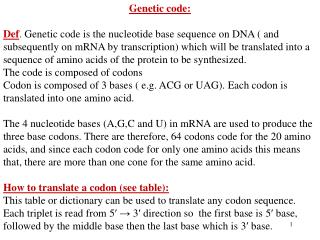
MOLECULAR BIOLOGY – DNA structure, genetic code Details in later lectures The Central Dogma of Molecular Biology - Francis Crick 1958 INFORMATION The genetic flow of information in a cell starts with DNA ‘instructions’ and passes through RNA ‘intermediates’ that dictate the synthesis of ‘functional’ protein BUT WHAT IS THE CODE BEHIND THIS TRANSFER OF GENETIC INFORMATION ?
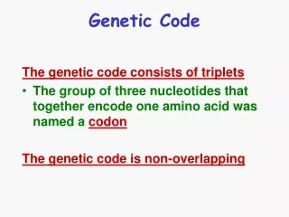
MOLECULAR BIOLOGY – DNA structure, genetic code Marshall Nirenberg & Heinrich Matthaei Synthetic poly-uracil RNA Observe if the radioactively labelled amino acid would be incorporated into protein? Lysed E-coli cell lysate (protein synthesis apparatus intact) Cracking the Genetic Code Proteins consist of 20 different amino acids whereas DNA/ RNA have only 4 different nucleotides (Uracil, replacing T in RNA): If a sequence of 2 nucleotides encoded a single amino acid the code could only accommodate 16 amino acids (i.e. 42) however a triplet nucleotide could code for potentially up to 64 amino acids (43) + 1 radiolabelled amino acid + 19 unlabelled amino acids Only when using labelled phenylalanine did the poly-uracil RNA lead to the production of radioactive protein The genetic code for the incorporation of phenylalanine into proteins had been cracked
MOLECULAR BIOLOGY – DNA structure, genetic code Three codons do not lead to incorporation of any amino acids - play role in terminating protein synthesis N.B. that the genetic code is largely redundant with most amino acids having more than one codon Methionine and tryptophan only have one codon The Genetic Code Similar experiments identified the other three letter ‘codons’ found in messenger RNAs (mRNAs) responsible for the incorporation of the remaining amino acids into protein - Nobel Prize of 1968
MOLECULAR BIOLOGY – DNA structure, genetic code Cracking the genetic code video/ tutorial http://bcs.whfreeman.com/thelifewire/content/chp12/1202002.html
MOLECULAR BIOLOGY – DNA structure, genetic code TRANSCRIPTION single stranded mRNA AUG GCU CCU UCU UCC AGA GGU GGC . . . . . . UAA TRANSLATION protein coding sequence or open reading frame MAPSSRGG….. Functional Protein DNA sequence driven Genetic Code of the Central Dogma double stranded DNA 5’ 3’ ATG GCT CCT TCT TCC AGA GGT GGC . . . . . . TAA TAC CGA GGA AGA AGG TCT CCA CCG . . . . . . ATT 3’ 5’ AUG UAA THE SEQUENCE OF SPECIFC NUCLEOTIDES IN DNA DICTATES THE SEQUENCE OF AMINO ACIDS IN THE FUNCTIONAL PROTEINS e.g. enzymes
MOLECULAR BIOLOGY – DNA structure, genetic code DNA STRUCTURE SHOULD FIT INTO 4 PRINCIPLES: 1. Provide a means for its own replication 2. Be able to encode the genetic information 3. Direct cell function 4. Accommodate changes caused by ‘mutations’
MOLECULAR BIOLOGY – DNA structure, genetic code 5’ 5’ 3’ 3’ ATG GCT CCT TCA TCC AGA GGT GGC . . . . . . TAA ATG GCT CCT TCT TCC AGA GGT GGC . . . . . . TAA TAC CGA GGA AGT AGG TCT CCA CCG . . . . . . ATT TAC CGA GGA AGA AGG TCT CCA CCG . . . . . . ATT 3’ 3’ 5’ 5’ AUG GCU CCU UCU UCC AGA GGU GGC . . . . . . UAA AUG GCU CCU UCA UCC AGA GGU GGC . . . . . . UAA Functional Protein Functional Protein Mutations are variations in the DNA sequence e.g. single base pair substitution double stranded DNA TRANSCRIPTION single stranded mRNA TRANSLATION AUG UAA protein coding sequence or open reading frame MAPSSRGG….. MAPSSSGG….. SUCH MUTATIONS CAN INFLUENCE THE FUNCTIONALITY OF THE PROTEIN e.g. changing which amino acid is incorporated
MOLECULAR BIOLOGY – DNA structure, genetic code Only 1.5% of the human DNA genome directly encodes amino acids for incorporation into proteins Most DNA is not coding for proteins !
MOLECULAR BIOLOGY – DNA structure, genetic code How is DNA organised in the cell? 600x Human DNA: 3 200 000 000 letters 200x 500-pagesbooks A single cells stretched out DNA = 1.8m
MOLECULAR BIOLOGY – DNA structure, genetic code Bacterial DNA ~ 1 mm long Most bacterial DNA exists in a covalently closed circular form 1000 x more than ~ 1 mm SUPERCOILING …thanks to mobiles no more twisted telephone cords!
MOLECULAR BIOLOGY – DNA structure, genetic code TOPOISOMERASES – enzymes that insert or remove supercoils Type I … break only one strand -> relaxing or twisting of the helix Type II … break both strands and pass another part of the double helix through the gap protein scaffold A typical bacterial chromosome consists of about 50 giant supercoiled loops of DNA
MOLECULAR BIOLOGY – DNA structure, genetic code Nucleosome H2A, H2B, H3, H4 80 bp 40 bp 80 bp 200 bp Eukaryotic DNA is complexed with HISTONE proteins that together form more and more ordered structures of CHROMATIN resulting in chromosomes
MOLECULAR BIOLOGY – DNA structure, genetic code Eukaryotic chromatin hierarcheal structure ‘beads on a string’ 30nm ‘solenoid fibre’ scaffold associated fibres Condensed chromosome Net result is that a eukaryotic (human) cell’s DNA is packaged into a mitotic chromosome 10,000 fold shorter than it extended length!
MOLECULAR BIOLOGY – DNA structure, genetic code SECOND SUMMARY – CRUCIAL KNOWLEDGE
MOLECULAR BIOLOGY – DNA structure, genetic code RNA T A C C G T T A G T T C A C G A T T A U G G C A A U C A A G U G C U A A A T G G C A A T C A A G T G C . . . . . . T A A CODING SEQUENCE TRANSCRIPTION DNA RNA RNA A U G G C A A U C A A G U G C U A A Met Ala Ile Lys Ala STOP START part of the chromosome where gene X is located double-strand DNA TRANSLATION Ribosomes, tRNAs (expanded later) PROPERLY FOLDED PROTEIN executes its function in cell The Central Dogma densely packed chromosome