Download
1 / 16
160 likes | 310 Views
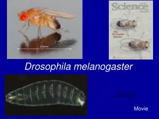
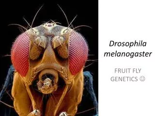
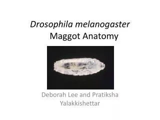
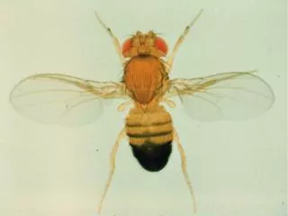
MOLECULAR ANALYSIS OF THE 2 ND FRAGMENT OF THE CINNABAR GENE IN DROSOPHILA MELANOGASTER. Dr Igor Donatovich Alexandrov Laboratory of Nuclear Problems JINR,Dubna, Moscow Region, Russia Nangamso Songca Walter Sisulu University Mthatha, Eastern Cape, South Africa. DROSOPHILA MELANOGASTER.

E N D
MOLECULAR ANALYSIS OF THE 2ND FRAGMENT OF THE CINNABAR GENE IN DROSOPHILA MELANOGASTER
Dr Igor Donatovich Alexandrov Laboratory of Nuclear Problems JINR,Dubna, Moscow Region, Russia Nangamso Songca Walter Sisulu University Mthatha, Eastern Cape, South Africa
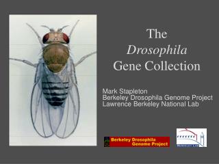
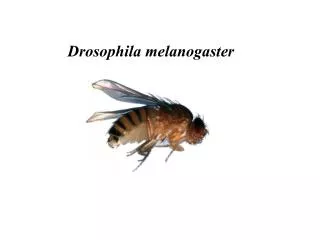
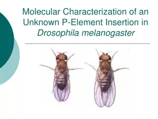
DROSOPHILA MELANOGASTER • It is commonly known as a fruit fly. • Well studied example, gene structure known and it is a commonly used model organism. • Has common principal DNA structure with humans. • Short life cycle (~15 days).
DROSOPHILA CONTIN’D • Recognizable match in gene diseases between humans and fruit flies.
RADIATION • Co 60 gamma rays and fission neutrons were used • Neutron radiation is used to treat caner. • Occupational exposure to neutrons.
AIM & OBJECTIVES • To detect DNA alterations in gamma ray and neutron induced cinnabar point mutants. • To improve the assessment of potential genetic risks resulting from exposure to neutrons.
METHODS • DNA isolation was performed using the Genomic DNA Isolation System. • The polymerase chain reaction (PCR) is a technique for the in vitro amplification of specific sequences of DNA. • PCR allows the detection of different kinds of mutational changes within segments.
METHODS CONTIN’D • PCR has three major steps which are based on thermal cycling. • Denaturation. • Annealing • Elongation
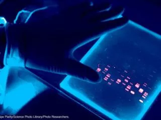
METHODS CONTIN’D • Agarose gel electrophoresis was used to separate mixed populations of DNA fragments.
VISUALIZATION • It was done using ethidium bromide under an ultraviolet light. • D 18(control), Cn116 (absent), Cn117, Cn 118, Marker, Cn123,Cn 142, Cn144,Cn175.
VISUALIZATION CONTIN’D • Ethidium bromide staining.
RESULTS AND CONCLUSION • There were 38 cinnabar point mutants that were studied 1 was exposed to neutrons only , 3 were exposed to neutrons and gamma rays simultanouesly, and 34 were exposed to gamma rays only. • Only 8.5% exhibited changes in DNA due to gamma rays. • There were no neutron radiation induced changes in all the mutants. • At the moment we can’t conclude that neutron are less effective than gamma rays this is a continuous project