Download

1 / 66
720 likes | 1.76k Views
Immunofluorescence and Confocal Microscopy. Dr. KW Chan. Part I: Immunofluorescence. Learning Objectives. To understand the working principles of immunofluorescence microscope To understand the difference between direct and indirect immunofluorescence

E N D
ImmunofluorescenceandConfocal Microscopy Dr. KW Chan
Learning Objectives • To understand the working principles of immunofluorescence microscope • To understand the difference between direct and indirect immunofluorescence • To know the current use of immunofluorescence studies in medicine
Principle of Fluorescence Microcopy Exciter filter
Principle of Fluorescence Microcopy Exciter filter Barrier filter
Fluorescence microscope • Light source is UV mercury vapor lamp • UV light is filtered to select excitation light to pass through • Excitation light is reflected by a dichroic mirror to strike on the specimen • Emission light passes through the dichroic mirror
Fluorescence microscope • Barrier filter blocks the excitation light amid the light path to visualization • Fluorescent labels are visulized against a dark background
Fluorescence microscope • The combination of exciter filter, dichroic mirror and barrier filter should be selected according to the fluorochrome label • The 3 components are usually built into a single module called the filter block
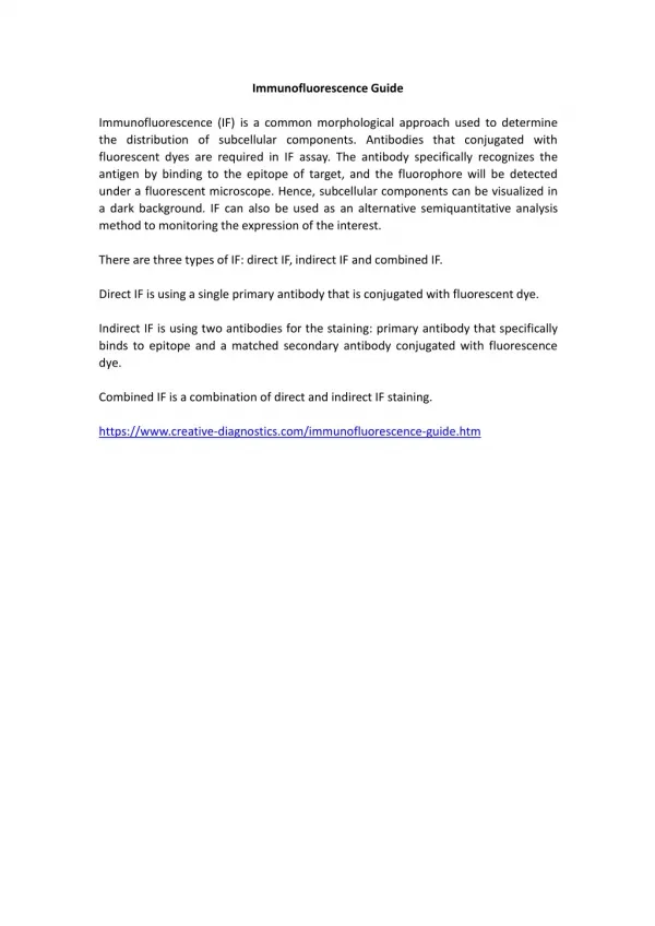
The basic principle of immunofluorescence • To use a fluorescent compound (usually fluorescein) to detect the binding of antigen and antibody. • The Ab is labeled with the fluorescent compound and its presence is detected using a fluorescence microscope. • Under a fluorescence microscope, fluorescein appears bright green wherever the binding occurred.
The basic principle of immunofluorescence • To use a fluorescent compound (usually fluorescein) to detect the binding of antigen and antibody • The Ab is labelled with the fluorescent compound • Under a fluorescence microscope, fluorescein appears bright green wherever the binding occurs
Using the fluorescence microscope • Select the correct filter block for the fluorescent compound • Fluorescence fades quickly under UV light; try to limit the time of exposure to UV as much as possible • Use high speed films for photography
Direct Immunofluorescence • The aim is to identify the presence and location of an antigen by the use of a fluorescent labelled specific antibody
Medical applications of direct IF • Renal diseases for evidence of immune deposition • Skin diseases for evidence of immune deposition • Detection of specific antigens, especially those of infective organisms
A section of kidney is placed on a slide; a fluorescein-labeled antiglobulin (specific for IgG, in this case) is added, then rinsed away • The presence of fluorescence in the glomeruli indicates that IgG was deposited prior to the biopsy • IgG is deposited in granular clumps along the capillary walls, enabling a diagnosis of membranous glomerulonephritis in this case
A section of kidney is placed on a slide; a fluorescein-labeled antiglobulin (specific for IgG, in this case) is added, then rinsed away • The presence of fluorescence in the glomeruli indicates that IgG was deposited prior to the biopsy • IgG is deposited in granular clumps along the capillary walls, enabling a diagnosis of membranous glomerulonephritis in this case
Direct Fluorescent Antibody Test for the Presence of Immunoglobulin Deposits in Skin IgG
A section of skin is placed on a slide; a fluorescein-labeled antiglobulin (specific for IgG, in this case) is added, then rinsed away • The presence of fluorescence in the upper layers of the epithelium indicates that IgG was deposited in this skin (prior to the biopsy) • The presence of immunoglobulins deposited around keratinocytes is consistent with a diagnosis of pemphigus
A section of skin is placed on a slide; a fluorescein-labeled antiglobulin (specific for IgG, in this case) is added, then rinsed away • The presence of fluorescence in the upper layers of the epithelium indicates that IgG was deposited in this skin (prior to the biopsy) • The presence of immunoglobulins deposited around keratinocytes is consistent with a diagnosis of pemphigus
Double labelling Lymphoid tissue: the two Ig light chains are separately labelled.
Indirect Immunofluorescence • The aim is to identify the presence of antigen specific antibodies in serum. The method is also be used to compare concentration of the antibodies in sera.
Indirect Immunofluorescence • A known antigen is placed on a slide; the patient's serum is added, then rinsed away. • A fluorescein-labeled antiglobulin is added, then rinsed away. • The presence of fluorescence over the antigen indicates the presence of antibodies to this antigen in the patient.
Diagnosis of Bacterial Diseases • Clostridial diseases (direct) • Brucella canis (indirect) • Afipia catei, cat scratch disease (indirect) • Borrelia burgdorferi (indirect) • Coxiella burnetii, Q Fever (indirect) • Rickettsia rickettsiae, Rocky Mountain Spotted Fever (indirect)
Diagnosis of Viral Diseases • rabies virus (direct) • bovine immunodeficiency-like virus (indirect) • canine coronavirus (indirect) • canine distemper (indirect) • feline infectious peritonitis (corona-) virus (direct) • porcine respiratory and reproductive syndrome (indirect)
Diagnosis of Protozoal Diseases • Babesia species (indirect) • Ehrlichia species (indirect) • Toxoplasma gondii (indirect) • Trypanosoma cruzi (indirect) • Cryptosporidia/Giardia (direct) • Encephalitozoon cuniculi (indirect) • Neosporum caninum (direct, indirect)
Some examples Indirect Immunofluorescence
Indirect Fluorescent Antibody Test for Antibodies to Toxoplasma gondii
Indirect Fluorescent Antibody Test for Antibodies to Toxoplasma gondii • Toxoplasma organisms are killed and placed on the slide; the patient’s serum is added, then washed away. • A fluorescein-labeled antiglobulin is added, then washed away. • The presence of the green fluorescence outlining the T. gondii organisms indicates the presence of antibodies in the patient's serum.
Indirect Fluorescent Antibody Test for Antibodies to Toxoplasma gondii
Immune-Mediated Disorders • antinuclear antibody (ANA) test (for diagnosis of systemic lupus erythematosus) • Direct fluorescent antibody test for deposition of Abs in tissues, e.g. kidney, skin
Indirect Fluorescent Antibody Test for Antinuclear Antibodies
Indirect Fluorescent Antibody Test for Antinuclear Antibodies • Cells from a cultured cell line are placed on a slide; the patient's serum is added, then rinsed away. • A fluorescein-labeled antiglobulin is added, then rinsed away. • The presence of fluorescence in the nucleus of these cells indicates the presence of antibodies to nuclear antigens in the patient.
Indirect Fluorescent Antibody Test for Antinuclear Antibodies
Advantage over Immunoperoxidase • Technically easier (fewer steps) • More sensitive results
Drawbacks • Microscope is more costly • Frozen sections preferred • Preparations need refrigeration • Preparations cannot be kept for too long • Quick fading of fluorescence under illumination (bleaching effect)
Learning Objectives • To understand the working principles of confocal scanning microscope • To know the current use of confocal scanning microscopy in medical science
Principles of confocal microscopy • a focused laser beam serves as a high intensity point source • light reflected or fluorescence emitted by the specimen is allowed to pass through a pinhole that filters light coming from outside (above and below) of the focal plane