Download
1 / 34
340 likes | 355 Views
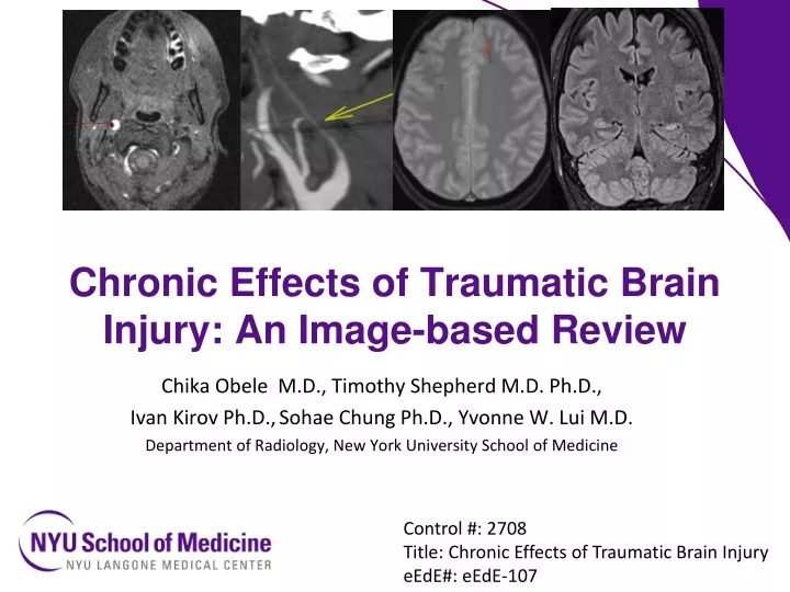
Chronic Effects of Traumatic Brain Injury: An Image-based Review. Chika Obele M.D., Timothy Shepherd M.D. Ph.D., Ivan Kirov Ph.D., Sohae Chung Ph.D., Yvonne W. Lui M.D. Department of Radiology, New York University School of Medicine. Control #: 2708

E N D
Chronic Effects of Traumatic Brain Injury: An Image-based Review Chika Obele M.D., Timothy Shepherd M.D. Ph.D., Ivan Kirov Ph.D.,Sohae Chung Ph.D., Yvonne W. Lui M.D. Department of Radiology, New York University School of Medicine Control #: 2708 Title: Chronic Effects of Traumatic Brain Injury eEdE#: eEdE-107
Background • Traumatic brain injury (TBI) is common (~1.9 million people annually in the U.S.) and can be devastating leading to hospitalization and fatalities. • With so may injury annually, aside from acute injury what are the chronic sequelae? • It is these that contribute to long-term morbidity and cost to society. • ~2% of the entire U.S. population lives with some long term disability due to TBI.
Educational Objective • To illustrate chronic sequelae of TBI with a focus on the role of imaging.
Imaging modalities CT is standard of care for assessment of acute head injury. MRI, however, is commonly used to evaluate long-term sequelae of TBI to assess: • Atrophy and gliosis • Potential epileptogenic foci • Small microhemorrhage • Brainstem and posterior fossa, typically difficult on CT. • A patient with unexplained persistent neurological deficits • And avoids ionizing radiation • research applications
Major secondary sequelae of TBI • Stroke / vascular • Post-traumatic epilepsy • Movement abnormality • Cognitive and behavioral difficulty • Hydrocephalus • Affective disorder
Vascular • There are a variety of vascular injuries that occur post-traumatically. • Can result in significant long-term morbidity. • Direct vascular injury • Ischemia (vasospasm, compression) • Hemorrhage • Venous infarct • AV Fistula • Pseudoaneurysm
Vascular Injury • 54yo F pedestrian with a complex skull base fracture (yellow) presented with massive epistaxis. Lateral projection from R CCA injection shows extravasation of contrast into the sphenoid sinus (red). Kelly clamp has been placed in the nasal cavity for packing. Courtesy Eytan Raz M.D.
Dissection • Classic imaging findings of carotid dissection in a patient with Horner’s syndrome and neck stiffness after TBI. • Crescentic mural thrombus on T1WI with fat saturation (red) and flame-shaped tapering on MIP CTA (yellow).
Stroke • Stroke is #1 cause of chronic disability in the U.S. • TBI known risk factor for stroke • Greater injury severity is associated with increased risk • e.g., TBI with fracture is associated with a 20-fold increase in stroke risk over TBI without fracture
Stroke • 65 year old male with a history of midline shift relating to post-traumatic hemorrhage resulted in bilateral anterior cerebral artery territory infarcts (red) • Chronic classic areas of inferior temporal contusion are also seen (light blue). • Major ACA (green) branches, seen here in a different patient, are susceptible to compromise as they can be pinned along the free edge of the falx in instances of substantial midline shift.
Stroke / Vascular • Axial FLAIR in a patient with traumatic left vertebral artery dissection shows PICA territory gliosis (yellow). • Time-of-flight MRA shows absent flow-related signal in the left vertebral artery (red).
Stroke • GRE T2* image in a 88-year-old man shows susceptibility in the sulci indicative of subarachnoid hemorrhage (yellow arrow) • Time of flight MRA MIP shows asymmetrically decreased flow-related signal in the left MCA (red) secondary to vasospasm.
Venous infarct • Rarely venous infarcts can complicate trauma. • 33yo with subdural hematoma (yellow), 2 weeks later developed massive left frontal lobe swelling (red) felt to be disproportionate to amount of initial contusional injury. • MIP TOF MRV shows compromise of a cortical vein (blue) along edge of decompressive craniectomy
Carotid Cavernous Fistula • Source data from CTA after trauma shows arterial phase contrast opacification of cavernous sinuses and superior ophthalmic veins bilaterally (red) • Note lack of equivalent contrast in the dural venous sinus (blue)
Dural AV Fistula • Occipital fracture is present (red) • Lateral projection selective L occipital artery injection shows dural arteriovenous fistula (yellow) with early venous drainage (blue).
Post-traumatic Seizure Disorder • Definition: recurrent seizures due to TBI • Risk factors: • penetrating injury • injury severity (GCS <10 in first 24 hours) • multiple contusions • >5mm midline shift • >24hrs loss of consciousness • dural penetration • prolonged amnesia • early post-traumatic seizures • injuries requiring surgical intervention • MRI can detect possible epileptogenic foci, such as areas of cortical contusion (red).
Post-traumatic seizure disorder • 47 year old female left temporal lobe injury after MVA 13 years ago • Has medically refractory posttraumatic seizure disorder • Underwent partial temporal lobectomy (blue) to resect gliotic brain though she developed left hippocampal sclerosis in the time since injury (red) and continued to suffer from seizures
Chronic Movement Disorders • Movement disorders are an uncommon complication of TBI • TBI is a known risk factor for Parkinson’s disease (44% increased risk in TBI patients) • Others: Choreoathetosis, hemiballismus, hyperreflexia. • Proposed mechanism is damage to deep gray matter (basal ganglia, thalamus), or disrupted basal ganglia / thalamocortical circuits. Ribbon cutting for the Muhammed Ali Parkinson’s Center in Phoenix, AZ http://mms.businesswire.com
Chronic Movement Disorders • Proposed mechanism is damage to deep gray matter (basal ganglia, thalamus), or disrupted basal ganglia / thalamocortical circuits • Hemorrhagic traumatic axonal injury shown here to the left basal ganglia (red) and right temporal white matter.
Cognition / Behavior • Patients may have problems with attention, memory, insight, judgment, language, communication and other executive functions. • Inferior frontal and temporal lobes are specifically at risk for contusion. • Frontal lobes play key role in behavior and higher-order cognition. • Frontal injury may lead to a clinical syndrome featuring poor impulse control, impaired attention, perseveration, and diminished divergent thinking.
Cognition / Behavior • Temporal lobe plays a key role in long-term memory, personality and affective behavior • Also susceptible to contusion (red) as it impacts the floor of the middle cranial fossa • Chronic injury results in gliosis and volume loss (blue)
Cognition / Behavior • In addition to focal contusion, traumatic axonal injury can cause widespread damage, affecting complex cognitive pathways. • Microhemorrhages are best detected using GRE T2*-weighted sequence (red) • SWI has higher sensitivity than standard GRE • Even subtle microhemorrhage such as seen here are clearly depicted (blue), in this patient with memory complaints after TBI. • Though subject to phase wrap artifact, phase map can be helpful to assess small lesions. • Here phase change is opposite that of the calcified choroid, consistent with blood products
Axonal Injury • It is known that foci of Traumatic Axonal Injury (TAI) may be seen on FLAIR and diffusion (red) without associated susceptibility (upper right) to suggest hemorrhage.
Axonal Injury • Much research points to diffusion abnormalities (MD, FA, kurtosis) predominantly shown using group analyses. • Current efforts use machine learning to identify injury in individual subjects • Pictured here are disorganized Tract-based density image (TDI) streamlines in a patient 2 years after moderate TBI (top image) compared with the usual symmetry seen in an age-matched control (bottom).
Neurodegenerative disorders • TBI increases relative risk for later development of a host of neurodegenerative disorders including: • Parkinson’s Disease (44% increased risk) • Alzheimer’s Dementia (>100% increased risk) • Chronic Traumatic Encephalopathy (CTE) is specifically associated with repetitive head trauma, recently described in professional contact sport athletes as a distinct pathologic entity
38 year old female • History of mild TBI now with persistent and progressive cognitive deficits • There is maintained overall brain volume with evidence of foci of TAI in the white matter.
Quantitative Volumetrics • The patient had asymmetric L hippocampal volume loss, with notably low asymmetry index compared with age-matched controls (arrow). • Hippocampal asymmetry is described in both Mild Cognitive Impairment (MCI) and Alzheimer’s dementia (AD).
FDG-PET • The patient demonstrated marked decrease in FDG uptake in a pattern reminiscent of Alzheimer’s Dementia (bitemporal and biparietal hypometabolism).
Hydrocephalus • Another factor that can contribute to cognitive decline, gait abnormalities, and shunt complications after TBI, shown here in a different patient with bifrontal cystic encephalomalacia after contusional injury.
Long-term brain structural changes • TBI associated not only with focal volume loss in areas of injury, but diffuse cortical atrophy. • Note the paucity of microvascular disease in this 54 year old patient with history of multiple head injuries and marked cerebral atrophy for age.
Conclusion • Though acute brain injury can be dramatic and life-threatening, it is the chronic sequelae of TBI that contribute to lifelong morbidity. • The main sequela span the gamut of neurological disease • vascular injuries leading to stroke, post-traumatic epilepsy, movement disorders, cognitive and behavioral changes, increased risk for neurodegenerative disorders • While CT is standard of care in assessing acute injury, MRI plays an important role in evaluating patients with TBI in the long-term especially those with persistent, unexplained symptoms.
References • Sosin, D.M., J.E. Sniezek, and D.J.Thurman, Incidence of mild and moderate brain injury in the United States, 1991. Brain Injury, 1996. 10(1): p.47-54. • Sosin, D.M., J.E. Sniezek, and R.J. Waxweller, Trends in Death Associated with Traumatic Brain Injury, 1979 through 1992 - Success and Failure. Jama-Journal of the American Medical Association, 1995. 273(22): p. 1778-1780. • Shively, S., et al., Dementia Resulting From Traumatic Brain Injury: What Is the Pathology? Arch Neurol, 2012: p. 1-7. • Max W, M.E., Rice DP, Head injuries: costs and consequences. J Head trauma Rehabil 1991. 6: p. 76-91 • Finkelstein, E., P.S. Corso, and T.R. Miller, The incidence and economic burden of injuries in the United States. 2006, Oxford ; New York: Oxford University Press. xiii, 187 • Latchaw, R.E., et al., Recommendations for imaging of acute ischemic stroke: a scientific statement from the American Heart Association. Stroke, 2009. 40(11): p. 3646-78. • Kelly, A.B., et al., Head trauma: comparison of MR and CT--experience in 100 patients. AJNR Am J Neuroradiol, 1988. 9(4): p. 699-708. • Lee, B. and A. Newberg, Neuroimaging in traumatic brain imaging. NeuroRx, 2005. 2(2): p. 372-83. • Englander, J., et al., Analyzing risk factors for late posttraumatic seizures: a prospective, multicenter investigation. Arch Phys Med Rehabil, 2003. 84(3): p. 365-73. • Chen, Y.H., J.H. Kang, and H.C. Lin, Patients with traumatic brain injury: population-based study suggests increased risk of stroke. Stroke, 2011. 42(10): p. 2733-9. Pitkanen, A. and T. Bolkvadze, Head Trauma and Epilepsy, in Jasper's Basic Mechanisms of the Epilepsies, J.L. Noebels, et al., Editors. 2012: Bethesda (MD). • Annegers, J.F., et al., Seizures after Head Trauma - a Population Study. Neurology, 1980. 30(7): p. 683-689. • Ates, O., et al., Post-traumatic early epilepsy in pediatric age group with emphasis on influential factors (vol 22, pg 279, 2006). Childs Nervous System, 2006. 22(10): p. 1376-1376. • Auterbach, M.D., et al., Treatment of Traumatic Brain Injury-Induced Dyskinesia With Tetrabenazine: A Case Report. Psychosomatics, 2014. • Gardner, R.C., et al., Traumatic brain injury in later life increases risk for Parkinson disease. Ann Neurol, 2015. 77(6): p. 987-95. • Schwarzbold, M., et al., Psychiatric disorders and traumatic brain injury. Neuropsychiatr Dis Treat, 2008. 4(4): p. 797-816. 90. • Deb, S., et al., Rate of psychiatric illness 1 year after traumatic brain injury. Am J Psychiatry, 1999. 156(3): p. 374-8. 91. . • Koponen, S., et al., Axis I and II psychiatric disorders after traumatic brain injury: a 30-year follow-up study. Am J Psychiatry, 2002. 159(8): p. 1315-21 Plassman etall Documented head injury in early adulthood and risk of Alzheimer's disease and other dementias. Neurology. 2000 Oct 24;55(8):1158-66. Gardner etal Traumatic brain injury in later life increases risk for Parkinson disease..Ann Neurol. 2015 Jun;77(6):987-95.
O-61 THANK YOU This work is supported in part by funding from the NIH/NINDS R01 NS039135